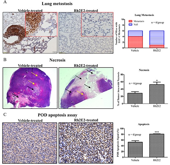
Figure 3. Immunohistochemical analysis of lung tumor tissues from Rh2E2-treated mice.

A. Rh2E2 suppressed the LLC-1 tumor metastasis in lung region of LLC-1 xenograft. Lung tissue sections from Rh2E2 or vehicle control-treated mice were stained with PCNA marker and its signal was visualized by DAB substrate followed by hematoxylin staining. Metastasized LLC-1 cancer cells with strong PCNA signals were visualized in lung tissues and captured from 6 animals of each group. Normal images, 10X magnifications; enlarged images, 40X magnification. Bar chart represented the number of mice with LLC-1 metastasis in lung region. B. Rh2E2 enhanced the necrotic areas in tumor tissues of LLC-1 xenograft mice. Tumor sections from Rh2E2 or vehicle control-treated mice were stained with hematoxylin and eosin. The necrotic area was shown in white color (black arrows), blood vessels were indicated as red color (yellow arrows). Bar chart represented the percentage of tumor area with necrosis. C. Rh2E2 increased the apoptotic cells in tumor tissue of LLC-1 xenograft. Tumor sections from Rh2E2 or vehicle control-treated mice were analyzed for apoptotic cells using POD kits followed by hematoxylin staining. Bar chart represented the percentage of apoptosis signal in tumor sections of LLC-1 xenograft.
