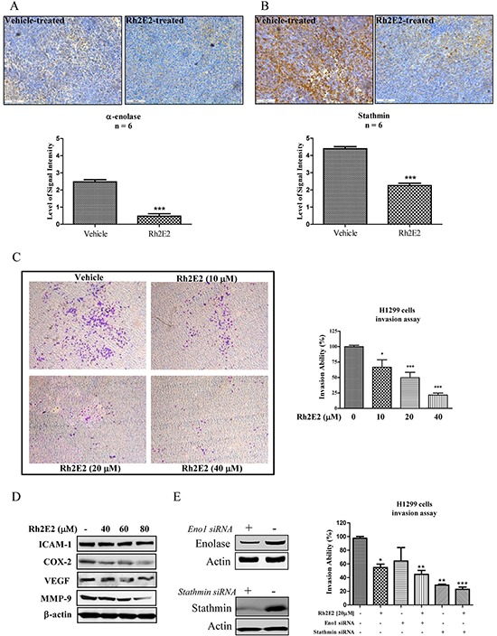
Figure 5. The role of α-enolase and stathmin in Rh2E2-inhibited cancer cells invasion.

A. Rh2E2 suppressed expression of α-enolase in tumor tissues of Rh2E2-treated LLC-1 xenograft mice. B. Rh2E2 suppressed the expression of stathmin in tumor tissues of Rh2E2-treated LLC-1 xenograft mice. α-enolase and stathmin staining images were representative of 5 tumor sections from 6 animals of each group. The level of signal intensity was scored from 1-5 (5 is maximum) and took average from five different views of each section taken in 20X magnifications. C. Rh2E2 dose-dependently inhibited the cell invasion ability of H1299 lung cancer cells. Images of invasive cells found in the lower layer of ECMatrixTM chamber were captured by Digital Camera under microscope with 40X magnifications. Bar chart represented the percentage of invasion ability of the stained invasive cells solute. D. Rh2E2 suppressed the expression markers for cell invasion and angiogenesis including ICAM-1, COX-2, VEGF and MMP-9. E. siRNA knockdown of α-enolase and stathmin potentiated the anti-invasive effect of Rh2E2. H1299 cancer cells were transfected with control or α-enolase and stathmin siRNA for 48 hours, the knockdown cells were then subjected to cell invasion assay using ECMatrix™ chamber followed by the treatment of Rh2E2. *P < 0.05, **P < 0.01 and ***P < 0.001 compared to medium control.
