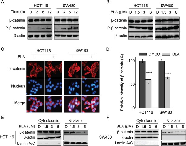
Figure 3. Bisleuconothine A promotes the phosphorylation and suppresses the nuclear translocation of β-catenin in colorectal cancer cells.

A. Western blot analysis of β-catenin and p-β-catenin (Ser33, Ser37 and Thr41) in HCT116 and SW480 cells treated with 3 μM of Bisleuconothine A (BLA) for 0 h, 3 h, 6 h and 12 h, respectively. β-actin was used as the loading control. B. Lysates from HCT116 and SW480 cells treated with DMSO (D), 1.5, 3 and 6 μM of Bisleuconothine A for 12 h were subjected to western blot analysis. The levels of β-catenin and p-β-catenin (Ser33, Ser37 and Thr41) were detected with β-actin used as the loading control. C. Representive figures of immunofluorescence staining. HCT116 and SW480 cells were incubated with or without Bisleuconothine A (3 μM). Distribution of β-catenin was examined by immunofluorescence staining. β-catenin and nucleus were recognized by the red and blue fluorescence, respectively. D. And the intensity of β-catenin in the nucleus of cells treated with DMSO and Bisleuconothine A was analyzed and quantified. All the values represent the mean ± S.D. (n=3). The significance was determined by Student's t test (***p<0.001 vs. control). HCT116 E. and SW480 F. cells were incubated with DMSO (D), 1.5, 3 and 6 μM of Bisleuconothine A for 12 h. The levels of β-catenin in cytoplasmic fraction and nucleic fraction of HCT116 and SW480 cell lysates were analyzed by Western Blot. β-actin and Lamin A/C is the loading control of cytoplasmic fraction and the nucleic fraction, respectively.
