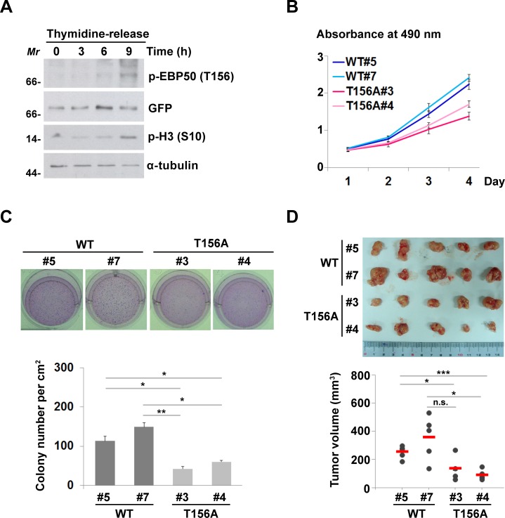
Figure 5. Cell cycle-dependent phosphorylation of EBP50 is crucial for cell proliferation and transformation.
A. HeLa cells expressing EGFP-EBP50 were synchronized at G1-S border using double thymidine block and released from the block for 0, 3, 6, or 9 h. Phosphorylation of EBP50 was assayed by Western blotting using phospho-T156 antibody. Phosphorylation of histone H3 at S10 was used as the mitotic marker. Molecular marker (Mr): kDa. B. Cell proliferation assayed using MTS reagent. Data are means ± SEM of 3 independent experiments. C. Photos and counting of cresyl violet-stained colonies formed in soft agar assay. D. Cells were injected subcutaneously into 8 weeks old NOD/SCID mice. Tumor volumes were measured in series for 4 weeks before the tumor were removed for photographing. Data are means ± SEM. * p < 0.05, ** p < 0.01, *** p < 0.001, n.s. not significant, Student's t-test.

