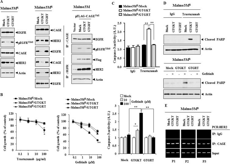
Figure 14. The inactivation of EGFR confers sensitivity to anti-cancer drugs and inhibits interactions of EGFR with CAGE and HER2.
(A) Malme3MR cells were treated with the indicated peptide (each at 10 μM) for 48 h. Cell lysates were then isolated and subjected to immunoprecipitation and immunoblot analysis (left panel). Malme3M cells were transfected with the indicated construct (each at 1 μg) along with the indicated peptide (10 μM). At 48 h after transfection, cell lysates were subjected to immunoblot analysis (right panel). (B) Malme3MR cells were treated with the indicated peptide (each at 10 μM) for 48 h. Cells were then treated with various concentrations of trastuzumab or gefitnib for 24 h, followed by MTT assays. *p < 0.05; **p < 0.005. (C) Malme3MR cells were treated with the indicated peptide (each at 10 μM) for 48 h. Cells were then treated with IgG (100 μg/ml), trastuzumab (100 μg/ml) or gefitinib (10, 100 μM) for 24 h, followed by caspase-3 activity assays. *p < 0.05; **p < 0.005. (D) Malme3MR cells were treated with the indicated peptide (each at 10 μM) for 48 h. Cells were then treated with IgG (100 μg/ml), trastuzumab (100 μg/ml) or gefitinib (10 μM) for 24 h, followed by immunoblot analysis. (E) Same as (A) except that ChIP assays were performed.

