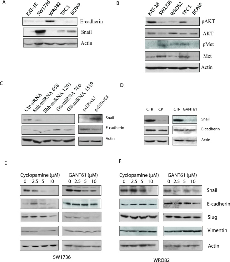
Figure 6. Effect of the Shh pathway on EMT.
A. & B. Cell lysates of KAT-18, SW1736, WRO82, TPC1, and BCPAP cell lines were analyzed for the expression of E-cadherin, Snail, and actin A. or pAKT, AKT, pMet, Met, and actin B. by Western blot. KAT-18 cells stably transfected with an expression vector encoding a control, Shh or Gli1 miRNA C. or transfected with pcDNA3.1 or pcDNA/Gli1 C. were analyzed for E-cadherin expression. (D-F) The effect of the Shh pathway inhibitors on E-cadherin expression. KAT-18 cells D. were treated with 0.5% DMSO as a vehicle control, cyclopamine (5 μM) or GANT61 (10 μM) for 72 hr. The cells were harvested and analyzed for E-cadherin and actin expression by Western blot with their specific antibodies. SW1736 E. and WRO82 F. cells were incubated in the presence of vehicle (0.5% DMSO) or the indicated concentrations of cyclopamine or GANT61 for 72 hr. The cells were harvested and analyzed for the expression of several genes involved in EMT.

