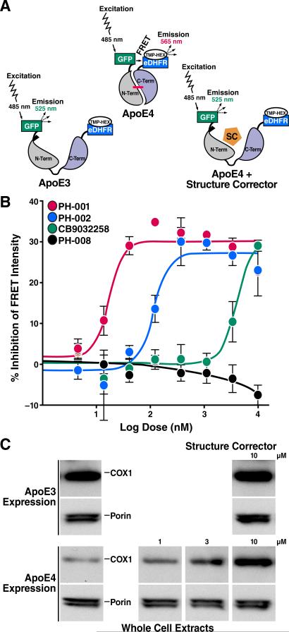
Figure 9. FRET Assay Identifies ApoE4 Structure Correctors.
(A) The FRET signal is generated between the donor and acceptor fluorophores conjugated to the N and C termini of apoE. ApoE4 gives a higher FRET signal because of the closeness of the N- and C-terminal domains. The apoE4 signal is reduced by the small-molecule apoE structure corrector (SC) by disrupting domain interaction (Chen et al., 2012).
(B) Dose-response analysis reveals the relative potencies of three active and one inactive structure corrector (Chen et al., 2012).
(C) Mitochondrial cytochrome c oxidase (COX1) is reduced in Neuro-2a cells expressing apoE4 compared with apoE3. A small-molecule structure corrector (CB9032258) corrects the COX1 deficiency in the apoE4-expressing cells, but does not significantly affect apoE3-expressing cells.
(A) and (B). Modified from Figure 2, Chen, H.-K., Liu, Z., Meyer-Franke, A., Brodbeck, J., Miranda, R.D., McGuire, J.G., Pleiss, M.A., Ji, Z.-S., Balestra, M.E., Walker, D.W., Xu, Q., Jeong, D.-e., Budamagunta, M.S., Voss, J.C., Freedman, S.B., Weisgraber, K.H., Huang, Y., Mahley, R.W. Small molecule structure correctors abolish detrimental effects of apolipoprotein E4 in cultured neurons. J. Biol. Chem. 2012; 287:5253–5266. © the American Society for Biochemistry and Molecular Biology.

