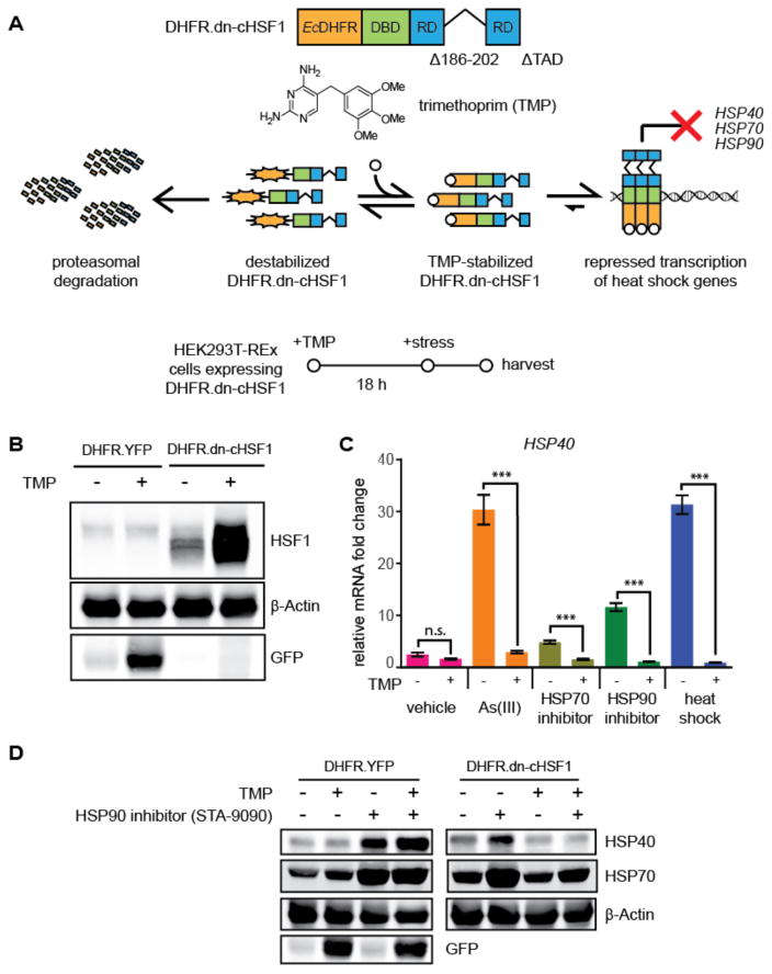
Figure 3. Application of destabilized domains to inhibit HSF1 in a small molecule-dependent manner.
(A) Model showing the TMP-dependent regulation of the DHFR.dn-cHSF1 fusion protein. The structure of TMP is shown.
(B) Immunoblot of HEK293T-REx cells expressing DHFR.YFP or DHFR.dn-cHSF1. TMP (10 μM) was added 18 h prior to harvest.
(C) qPCR analysis of HSP40 in HEK293T-REx cells expressing DHFR.dn-cHSF1. Vehicle (DMSO) or TMP (10 μM) was added for 12 h, followed by treatment with arsenite (100 μM; 6 h), HSP90 inhibitor STA-9090 (100 nM; 6 h), HSP70 inhibitor MAL3-101 (10 nM; 6 h) or heat shock (42 °C; 2 h). qPCR data are presented as fold-increase relative to vehicle-treated cells expressing DHFR.YFP. Error bars represent SEM from biological replicates (n = 3). ***p < 0.0001.

