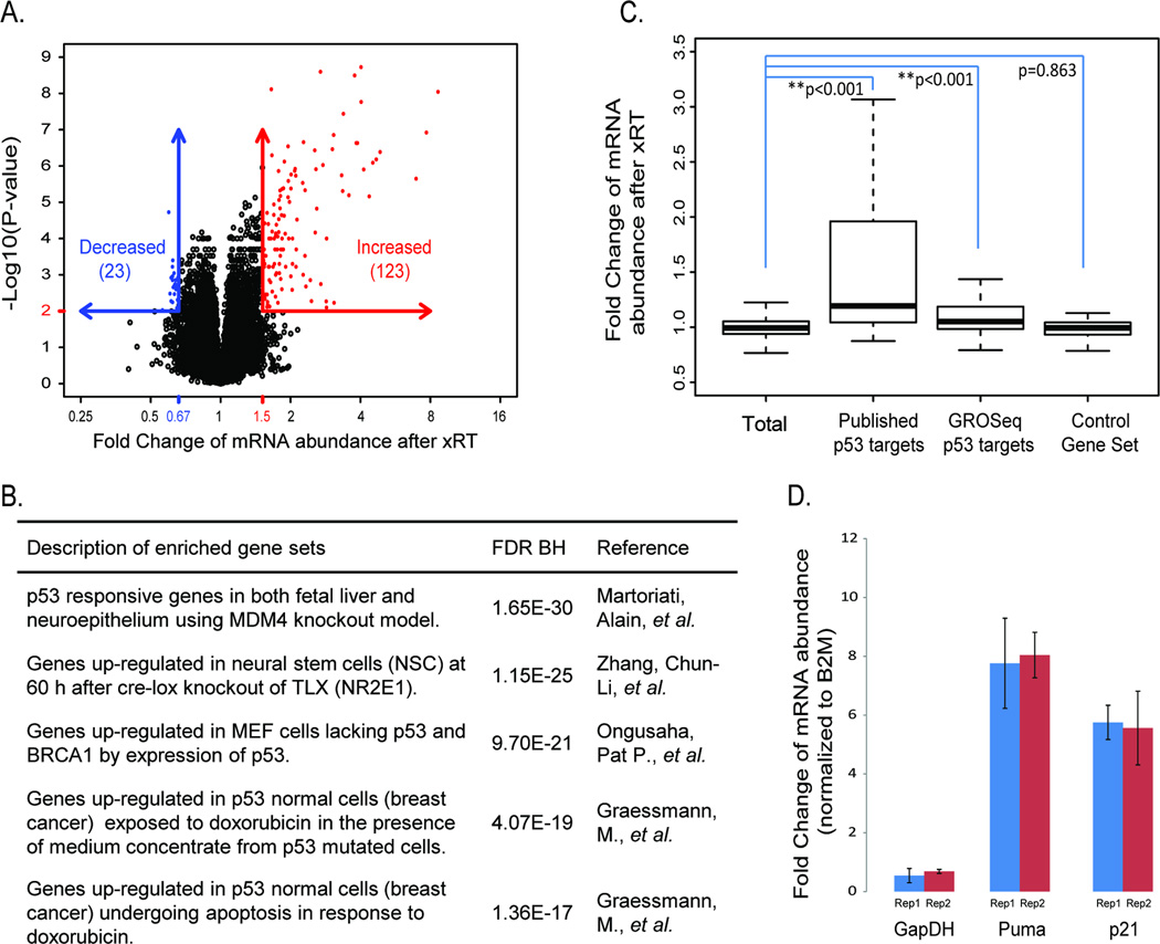
Figure 2. Increased expression of p53 targets mediating apoptosis and cell cycle arrest after xRT.
A) Quantification of mRNA abundance after xRT based on expression microarray data. B) The five most enriched gene sets from the genes with increased expression using GSEA analysis (see supplementary table 1). C) Comparison of mRNA abundance change after xRT among indicated gene sets (see supplementary table 2). P-values are estimated by permutation. D) Fold change in the abundance of mRNA for Puma, p21 and GAPDH when radiated and untreated M-Smo tumors are compared (n=3 for each group). Abundance of mRNA was measured by quantitative RT-PCR and normalized to the abundance of β2 Microglobulin (B2M) mRNA. Puma and p21 are induced by xRT, while GAPDH abundance is not significantly altered.

