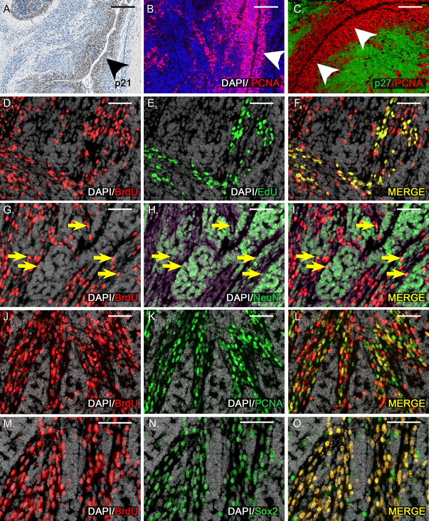
Figure 7. In apoptosis-deficient tumors, xRT fails to prevent the growth of perivascular stem cells that drive recurrence.
A) IHC demonstrates p21 up-regulation specifically in cells of the perivascular region (arrowhead) 4 hours after xRT. B) 48 hours after xRT, cells of the perivascular region (arrowhead) express proliferation marker PCNA. C) Double labeling with antibodies to PCNA and differentiation marker p27 demonstrates proliferation in the perivascular region (arrowheads) and differentiation further from the vessels. D–F) BrdU injected 5 days after xRT labels a subpopulation of tumor cells in a representative M-Smo;Baxfloxed tumor. EdU injection 2 hours prior to harvest labels an overlapping subpopulation. All EdU+ cells were BrdU+, indicating that cells proliferating 7 days after xRT descend from cells proliferating 2 days earlier. Some BrdU+ cells were EdU- consistent with a portion of BrdU+ cells having left the cell cycle. G–I) In the same tumor, a portion of BrdU+ cells express NeuN (yellow arrows), consistent with neuronal differentiation. J–L) In the same tumor, numerous BrdU+ cells are PCNA+, indicating continued proliferation. M–O) BrdU+ cells were predominantly Sox2+, consistent with a stem cell phenotype. Scale bars: 100 µm (A–C), 50 µm (D–O).

