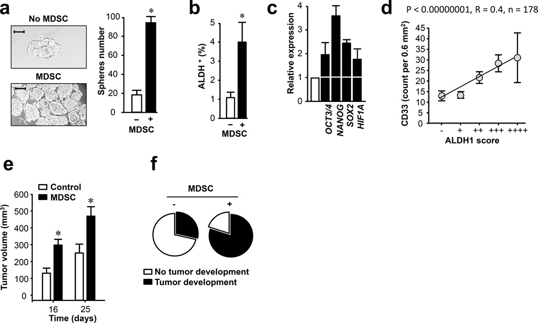
Figure 3. MDSCs promote and correlate human breast cancer stem cells.
(a) Human MDSCs increased breast cancer sphere formation. Human MCF-7 breast cancer cells were cultured with MDSCs in sphere forming condition (24). Numbers of spheres are expressed as Mean ± SEM, n = 4, MDSCs derived from 3 different patients. *, P<0.05, Wilcoxon pair test.
(b) Human MDSCs increased ALDH+ breast cancer stem cells. MCF-7 breast cancer cells and MDSCs were co-cultured in sphere forming condition (24). ALDH+ cells were determined by FACS gated on CD33− tumor cells. Results are expressed as Mean ± SEM, triplicates with MDSCs derived from 3 different patients. *, P<0.05, Wilcoxon pair test.
(c) Human MDSCs stimulated human breast cancer stem cell core gene transcripts. MCF-7 cells were co-cultured with primary MDSCs in transwell for 48 hours. Stem cell core genes quantified by real-time PCR. Results are expressed as the mean relative values ± SD. Experiments were triplicates with MDSCs from 3 different patients.
(d) Relationship between MDSCs and ALDH-1+ cancer stem cells in breast cancer tissues. CD33+ cells and ALDH-1+ tumor cells were evaluated by immunohistochemistry staining in tissues obtained from patients of cohorts 1 and 2. CD33+ cells were quantified as the numbers of CD33+ cells/0.6mm2 in TMAs. ALDH-1+ cells were scored from 0 to 4 based on the intensity of ALDH-1 expression (see Methods). Their correlation was analyzed by Pearson correlation. N = 178; P < 0.00000001, R = 0.4.
(e) Effects of MDSCs on human MCF-7 breast tumor growth in NSG mice. Human breast cancer MCF7 cells were mixed with MDSCs and inoculated subcutaneously into NSG mice supplied with E2 (estradiol-17β pellet). Tumor growth was monitored. N = 5/group; *, P < 0.05 (Wilcoxon pair test).
(f). Effects of MDSCs on human MCF-7 incidences in NSG mice. 105 MCF7 cells were mixed with MDSCs and inoculated subcutaneously into NSG mice supplied with E2 (estradiol-17β pellet). Tumor incidence was monitored for 16 days. N = 5/group.

