Figure 2.
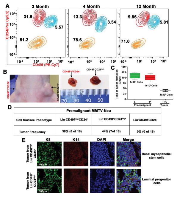
Distinct myoepithelial stem cells and luminal progenitor cells represent the cells of origin. A, Representative contour plot of FACS gating shows an abnormal increase in luminal cells and shifting of myoepithelial stem cells toward luminal cells in 3-month-old MMTV-Neu premalignant mice. In contrast, 4- and 12-month-old mice show normal level of stem and luminal cells (n=3 mice in each). B, Tumors in Balb/c mice, driven by basal myoepithelial stem cells and luminal progenitor cells, which were derived from premalignant MMTV-Neu mice (n=3 mice). C, Tumor-forming potential of stem cells and luminal cells from premalignant MMTV-Neu mice, and tumor-propagating cells from MMTV-Neu mouse tumor tissue (n=3 mice). D, Frequency of tumor formation by different cell types, derived from MMTV-Neu premalignant mice. E, Representative confocal images (10x) of keratin 8 (green), keratin14 (red) and DAPI (blue) staining for sections of tumor tissues derived from transformed stem cells and luminal cells (n=3 mice). Scale bars are 100 μm.

