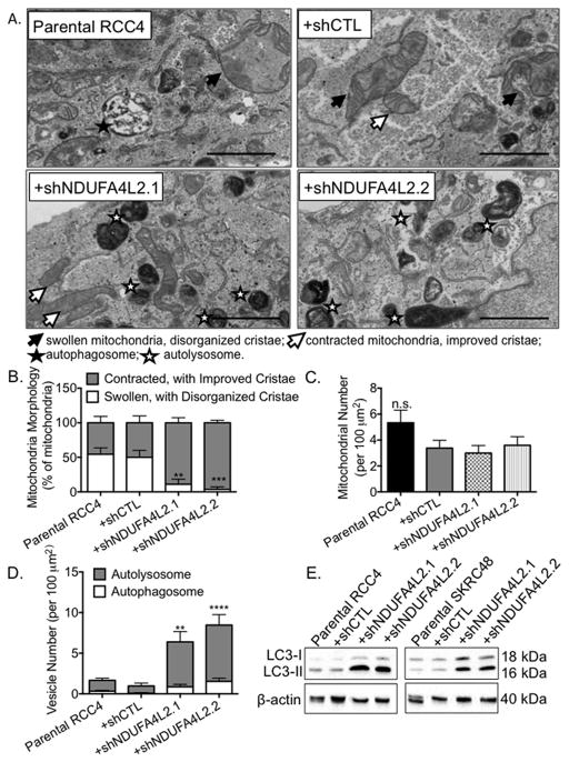
Figure 5. NDUFA4L2 knockdown improves mitochondrial morphology and induces autophagy in RCC4 cells.
(A) Representative images from transmission electron microscopy of RCC4 parental cells and cells infected with the indicated shRNA constructs for 24 hours followed by puromycin for 96 hours. Filled arrows point to swollen mitochondria with disorganized cristae, open arrows point to contracted mitochondria with improved cristae, filled stars indicate autophagosomes, and open stars indicate autolysosomes. Magnification, 25000 x. Scale bar on the bottom left represents 2 μm. (B) Quantification of the percentages of swollen mitochondria with disorganized cristae compared to the percentages of contracted mitochondria with improved cristae. (C) Quantification of the number of mitochondria per 100 μm2. (D) Quantitation of the number of autophagosomes and autolysosomes per 100 μm2 in RCC4 cells. (E) Western blot analysis of LC3 in RCC4 and SKRC48 cell lines following 24 hours of shRNA treatment, 96 hours of puromycin selection, and a 5 day, untreated, incubation period. β-actin was used as loading control. Quantification of TEM data was performed in a blinded manner. Data were collected from three images from five different cells (15 images/cell type) and graphed as mean ± SEM. ** p < 0.01; *** p< 0.005; **** p < 0.0001; n.s. p>0.05.

