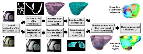
Fig. 3.
Block diagram of model generation, and execution of electrophysiological simulations, for the evaluation of our infarct segmentation method. the letter(s) in parenthesis in a block refer(s) to the corresponding subsection(s), of Section II, where the processing in the block is described. From each LGE-CMR image, two ventricular models, one incorporating infarct geometry reconstructed from manual segmentation, and the other with infarct geometry reconstructed from computed segmentation, were generated. Outcomes of electrophysiological simulations with the two models were then compared.

