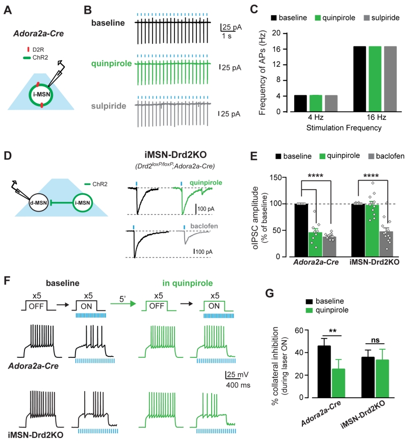
Figure 3. Quinpirole inhibits collateral GABA transmission to enhance dMSN excitability via D2Rs in iMSNs.
A, Schematic of experimental configuration. B, Traces of cell-attached recordings from iMSNs expressing ChR2 showing high fidelity of AP firing during opto-stimulation at baseline (black) and in quinpirole (1mM, green) followed by sulpiride application (1mM, grey). C, AP firing frequency during trains of light pulses at 4 and 16 Hz under each drug condition (n = 5-8). D, Schematic of experimental configuration and oIPSC traces recorded from dMSNs in iMSN-Drd2KO mice at baseline (black) and during quinpirole (1 μM, green) and baclofen (5 μM, grey) application. E, oIPSC amplitude as percent of baseline during quinpirole and baclofen in control Adora2a-Cre mice (n = 6-11) and iMSN-Drd2KO (n = 8-13). F, Top, Current pulses delivered to dMSNs before and during opto-stimulation of iMSN axon collaterals. Bottom, AP traces recorded from dMSNs in control Adora2a-Cre and iMSN-Drd2KO mice before (OFF) and during (ON) opto-stimulation. G, Percent inhibition of AP firing by activation of iMSN collateral transmission at baseline and in quinpirole for Adora2a-Cre (n = 12) and iMSN-Drd2KO (n = 11). ns = not significant, **p < 0.01, ****p < 0.0001. All data expressed as mean ± s.e.m.

