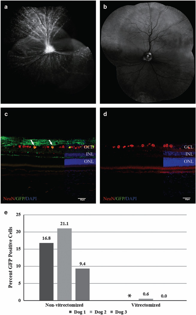
Figure 1.
Comparison of ganglion cell layer transduction efficiency of non-vitrectomized and vitrectomized eyes following IVT of AAV2 (triple Y-F+T-V). Representative confocal scanning laser ophthalmoscopy images (a, b) obtained at 5 weeks post injection demonstrate widespread fluorescence in non-vitrectomized eye (a) versus fluorescence limited to the optic nerve head of the vitrectomized eye. (b) The low level of background autofluorescence in the superior retina of image (b) is the expected appearance of the canine tapetum. Immunohistochemical labeling of retinal cryosections with NeuN antibody (c, d) demonstrating a higher number of cells colabeling with GFP in non-vitrectomized eye (c, white arrows) compared with absent transduction in vitrectomized eye (d). (e) Overall ganglion cell layer transduction efficiency for non-vitrectomized eyes and vitrectomized eyes 6 weeks post IVT. Non-vitrectomized eyes had significantly higher levels of ganglion cell layer transduction (P=0.04; unpaired t-test). DAPI, 4',6-diamidino-2-phenylindole; GCL, ganglion cell layer; GFP, green fluorescent protein; INL, inner nuclear layer; IVT, intravitreal injection; ONL, outer nuclear layer. Scale bar, 50 µm. *There is no transduction efficiency calculation for the vitrectomized eye of Dog 1 due to loss of sample for histopathological analysis.

