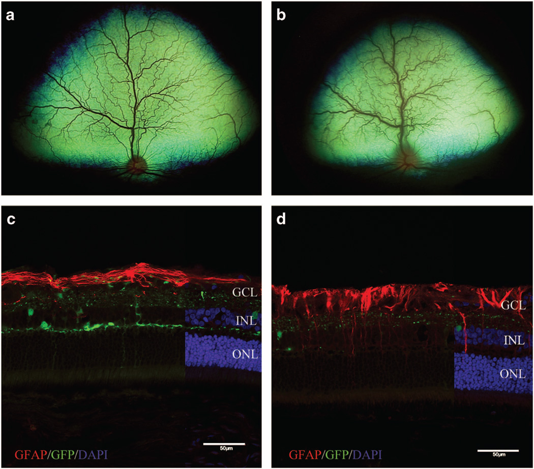
Figure 2.
White-light fundoscopic imaging showing the normal fundus of the non-vitrectomized eye of Dog 1 at 4 weeks post IVT (a) and acute retinitis that developed during week 6 (b). Fluorescent microscopic images of retinas from non-vitrectomized eyes labeled with GFAP shown in c and d to demonstrate glial activation of Müller cells as a result of inflammation. (c) Retina from Dog 2 without gross evidence of inflammation, showing expected strong GFAP labeling limited predominantly to the nerve fiber layer. (d) Retina from Dog 3 with chronic inflammation, showing GFAP labeling extending from the nerve fiber layer into the inner and outer retinal layers. DAPI, 4',6-diamidino-2-phenylindole; GCL, ganglion cell layer; GFP, green fluorescent protein; INL, inner nuclear layer; IVT, intravitreal injection; ONL, outer nuclear layer. Scale bar, 50 µm.

