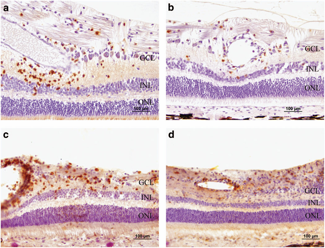
Figure 4.
Photomicrographs characterizing the cellular component of the retinal inflammatory response from vitrectomized eye of Dog 1 (a, b) and non-vitrectomized eye of Dog 3 (c, d) through the use of CD labeling. Positive immunolabeling of mononuclear inflammatory cells with CD20 (a) and CD3 (b) demonstrates a mixed response involving both B cells and T cells, respectively. Positive immunolabeling of inflammatory cells with CD4 (c) and CD8 (d) demonstrates that both T-helper and cytotoxic T cells, respectively, were present. GCL, ganglion cell layer; INL, inner nuclear layer; ONL, outer nuclear layer. Scale bar, 100 µm.

