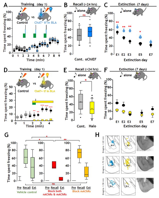
FIGURE 2. Optogenetic manipulation of cholinergic signaling in BLA regulates fear and extinction learning Effects of activating cholinergic terminal fields in BLA during tone-shock fear-conditioning (A – C).
A: Training - Top: Schematic of training with optogenetic stimulation of oChIEF expressing cholinergic terminal fields in BLA. On the training day tone/shock pairs (green block/black line) were presented 3 times at 2 min intervals. At 27.5 s after each tone onset, mice in the opto group received 100 × 5 msec 473 nm optical pulses (20 Hz, 10 mW/mm2) to the dorsal surface of the BLA. Mice in control group were implanted with light guides but received no optical stimulation. Bottom: Plot of percent time spent freezing during training (opto-stimulation group: grey/blue boxes; control group: black boxes).
B: Recall - Top: Schematic of paradigm for test of recall. Twenty four hours after training mice were exposed to the same 30 s tone as on the training day and recall was measured as the time spent freezing. Bottom: There was no significant difference in recall between Control (Cont.) and oChIEF activated groups. Data are presented in box plot format to show the full range and distribution of the data; the boundaries of the box demarcate 75% of the data points, the whiskers delineate the 95% range and additional values outside of the 95% range are shown. The horizontal bar indicates the population median.
C: Extinction – Top: Schematic of paradigm for extinction training. Extinction learning involved exposing the mice to the same 30 s tone 10 times at pseudo-random intervals. Bottom: Retention of extinction learning was tested 24 hours after each extinction training session by measuring the time spent freezing in response to the first 30 s tone (mean ± SEM). Extinction training and test of recall were repeated daily for 7 days. Significant differences in freezing as a function of days of extinction learning were found between control and oChIEF groups at all days except on day 7.
Inhibition of cholinergic terminal fields: effects on recall and extinction (D – F)
D: Training – Top: Schematic of training paradigm with optogenetic inhibition of cholinergic terminal fields in BLA. On the training day, mice were exposed to one 30 s, 5 kHz, 80 dB tone (green block) co-terminating with a 2 s, 0.8 mA foot shock (black line). At the tone onset, mice in the Halo group (grey/yellow; n=22), received 2.5 min of continuous optical stimulation (12–15 mW/mm2, 590 nm, yellow block) of the dorsal surface of the BLA. Control mice (black, n = 18) received no optical stimulation. Bottom: Plot of time spent freezing over the 4 min of training with or without optogenetic inhibition. Prior to the cue x optogenetic pairing there was no significant difference in time spent freezing between groups. Immediately after the onset of optical stimulation, a highly significant difference in freezing was found (mean ±SEM; p < 0.001).
E: Recall - Top: Schematic of test of recall (as in 2B). Recall was measured as the time spent freezing to the tone. Bottom: There was a statistically significant difference in the extent of Recall between Control and Halo trained groups (p = 0.018).
F: Extinction - Top: Schematic of paradigm for extinction training. Extinction training and test of retention of extinction learning were performed as described above. Bottom: Overall there was no significant difference in the retention of extinction learning between groups.
G: Effect of intra-BLA infusion of cholinergic antagonists on fear conditioning and retention of extinction learning. To test the contribution of acetylcholine receptors to fear conditioning and retention of extinction learning we compared the behavior of mice with bi- lateral, intra BLA injections of saline alone (Vehicle Control) vs those injected with blocking concentrations of both nicotinic and muscarinic acetylcholine receptor antagonists (mecamylamine 20uM + atropine 1uM) vs those injected with blocking concentrations of atropine alone (1uM). Blocking concentrations of both nAChR and mAChR antagonists, but not mAChR antagonists alone, reduced recall (saline n=17, atropine & mecamylamine n=8, atropine n=9; one way ANOVA with Bonferroni correction: F(2,31)=6.935, p = 0.003 for group effect; saline recall vs atropine & mecamylamine recall, p=0.017; saline recall vs atropine alone recall, p=0.797; atropine alone recall vs atropine & mecamylamine recall, p=0.003). There was significantly greater retention of extinction learning in the group administered both atropine & mecamylamine as compared to vehicle control, while the degree of retention of extinction learning was unaffected by atropine (one way ANOVA on ranks; H=6.417, df=2, p=0.04; control vs mixed antagonists p<0.05).
H: Location of fiberoptic track terminations in fixed sections in oChIEF expressing (blue dots), or Halo–expressing mice (yellow dots). See Fig S2, Supplemental Video and Table S1.

