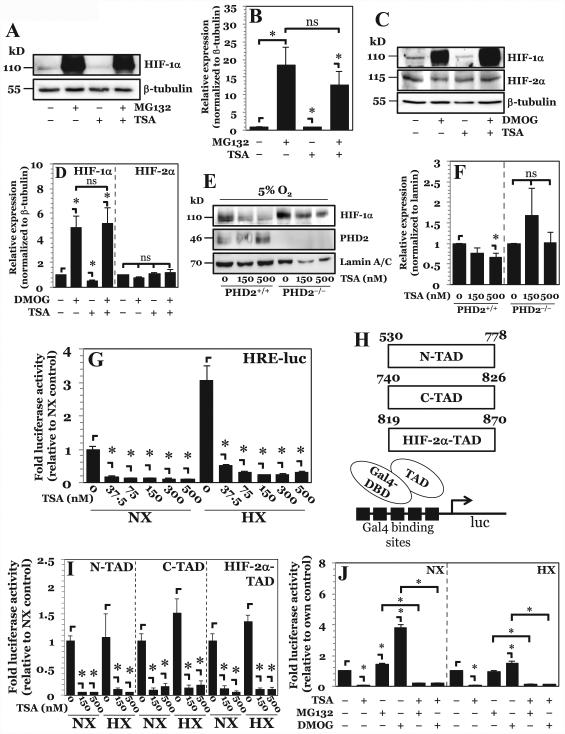
Figure 2. Higher-dose TSA treatment results in increased HIF-1α turnover through PHD-26S proteasome pathway while lower-dose TSA treatment independently decreases HIF-1α activity.
A, B) Western blot analysis of HIF-1α (A) and corresponding densitometric quantification of at least 3 independent experiments (B) after treatment of NP cells with either proteasomal inhibitor MG132 (10 μM) or TSA (500 nM), or both. Decrease in HIF-1α protein seen with higher-dose TSA treatment is rescued by proteasomal inhibition. C, D) Western blot analysis (C) and corresponding densitometry (D) of HIF-1α in NP cells after exposure to PHD inhibitor dimethyloxalylglycine (DMOG, 2 mM) TSA (500 nM), or both. Decrease in HIF-1α protein levels seen with higher-dose TSA treatment is rescued by PHD inhibition. E, F) Western blot (E) and corresponding densitometry of at least 3 independent blots (F) of HIF-1α in PHD2+/+ and PHD2−/− MEFs after exposure to TSA. HIF-1α shows no degradation following higher-dose TSA treatment in PHD2−/− cells. G) HRE-luciferase reporter activity under both normoxia and hypoxia following treatment with increasing doses of TSA. HRE activity decreases with all doses of TSA irrespective of oxygen tension. H) Schematic of Gal-4-TAD constructs and the assay system used for studies described in (I). I) HIF-1α-N-TAD, -C-TAD, and HIF-2α-TAD activity under normoxia and hypoxia after TSA treatment. Both lower dose and higher dose TSA decrease TAD activity under both normoxia and hypoxia. J) HRE-luc activity in NP cells under NX or HX after treatment with MG132, DMOG, and TSA. Data is represented as mean ± S.E. of three independent experiments performed in triplicate (n = 3); *, p < 0.05.

