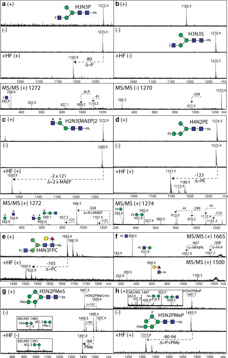
Fig. 3.
Comparative mass spectrometry of N-glycans modified by phosphoesters and sulphate. Example positive and negative ion mode MALDI-TOF MS and MS/MS (also after hydrofluoric acid treatment) of N-glycans from invertebrate and protist organisms. (a-c) Comparison of the positive (+) and negative (−) ion mode MALDI-TOF MS spectra before and after hydrofluoric acid (HF) treatment of isobaric N-glycans modified with either phosphate (P), sulphate (S) or methylaminoethylphosphonate (MAEP or *) from two marine organisms together with a relevant MS/MS spectrum of either the m/z 1270 or 1272 [M-H]− or [M + H]+ quasimolecular ions. (d) Positive (+) and negative (−) ion mode MALDI-TOF MS spectra before and after hydrofluoric acid treatment of a Trichomonas vaginalis N-glycan (Tv2 strain) with the corresponding MS/MS spectrum of m/z 1274. (e, f) Positive ion mode MALDI-TOF MS and MS/MS of a nematode N-glycan modified with phosphorylcholine (PC) before and after hydrofluoric acid treatment. (g, h) Comparison of the positive (+) and negative (−) ion mode MALDI-TOF MS spectra before and after hydrofluoric acid (HF) treatment of isobaric N-glycans modified with methylphosphate (PMe) and phosphate or sulphate from Dictyostelium with relevant MS/MS spectra of either the m/z 1485 or 1487 quasimolecular ions. Further structural details were in each case proven by enzymatic digestion. The data in a are unpublished, whereas those in panels b-h underlie assignments in previous publications on Volvarina rubella (b and c; marine snail), Trichomonas vaginalis (d; protist), Caenorhabditis elegans (e and f; nematode) and Dictyostelium discoideum (g and h; slime mould) [4, 14, 16, 74]

