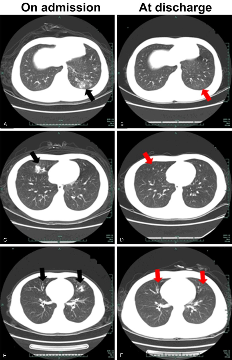
Figure 1.

Chest computer tomography (CT) scans in 3 hospitalized patients with HAdV7 infection on admission (A, C, E) and at discharge (B, D, F). Three pulmonary infection pattern were showed: high density mottling or patchy shadows (black arrows) in the left lobe (A), in the right lobe (C), and in bilateral lungs (E).
