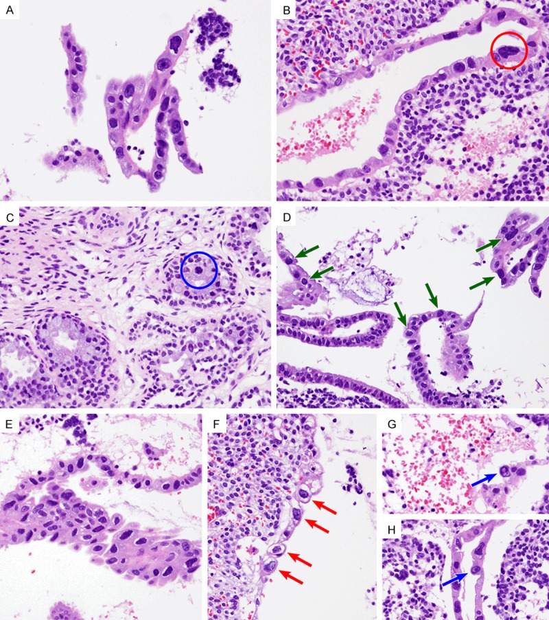
Figure 1.

Histopathological findings. A. Detached strips of endometrial glandular epithelium revealed atypical cells showing abundant, amphophilic, partly vacuolized cytoplasm and enlarged nuclei with irregular outlines and hyperchromasia. The nuclear/cytoplasmic ratio was not increased, because the quantity of cytoplasm was also increased. A few conspicuous nucleoli were identified. B. A bizarre nucleus (red circle) was approximately five times larger compared to its normal counterpart. C. An endocervical gland had a few large, hyperchromatic nuclei (blue circle). D and E. Variable distribution of the atypical cells was observed. D. In some areas, atypical cells showing variable degrees of nuclear pleomorphism were distributed in a patchy fashion (green arrows). E. In other areas, pseudostratified atypical cells with a degenerative-looking chromatic pattern showed architectural disarray. F-H. Intracytoplasmic vacuolar change (red arrows) and/or intranuclear microvesiculation (blue arrows) raise the suspicion of viral-induced cytopathic effect.
