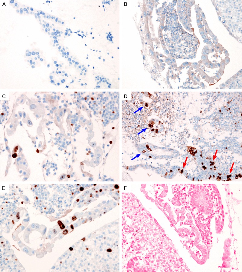
Figure 2.

Immunohistochemical findings. Lack of (A) HSV and (B) CMV immunoreactivity excluded the possibility of a viral-induced cytopathic effect. (C) Patchy nuclear staining for p53 indicated the absence of the TP53 mutation. (D) The patchy expression pattern of p16 expression in atypical cells (blue arrows) was the same as that in normal endometrial glandular epithelium (red arrows). (E) Ki-67 proliferation index was low (less than 5%) in the atypical epithelial cells. (F) EBER-ISH did not reveal EBV infection in the atypical epithelial cells.
