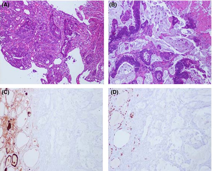
Figure 1.

Histological and immunohistological characterization of thyroid metastasis. (A) H&E of moderately differentiated primary tumor with mucinous features; (B) H&E of metastatic thyroid lesion reveals poorly differentiated colonic adenocarcinoma with desmoplastic features in a background of extracellular mucin; (C) Immunohistochemistry for thyroglobulin (TG) demonstrates staining of the resident thyroid tissue (brown) and no staining of metastatic tumor cells; (D) Immunohistochemistry for thyroid transcription factor‐1 (TTF‐1) demonstrates staining of the resident thyroid tissue (brown) and no staining of metastatic tumor cells.
