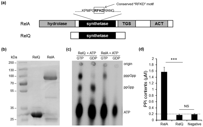
Figure 1. Expression and activity analysis of RelA and RelQ proteins of S. suis.
(a) Domain structures of RelA and RelQ of S. suis. The RXKD motif was labeled in the box, TGS, conserved domain on RSH C-terminal domain for uncharged ACP binding in E. coli, ACT, a conserved regulatory domain on RSH C-terminal domain. (b) SDS-PAGE analysis of purified recombinant RelA and RelQ proteins. (c) Thin-layer chromatography (TLC) analysis of the (p)ppGpp synthesis activity of the recombinant RelA and RelQ. Purified RelA or RelQ protein is assayed for (p)ppGpp synthetase activity in the presence of [γ-P32]-ATP, 2 mM ATP, and either 1.3 mM GTP or GDP. Reaction mixtures were analyzed by TLC and autoradiography as described in Materials and Methods. (d) Hydrolase activity assays of recombinant RelA and RelQ by measuring the ppGpp hydrolysis product, PPi.

