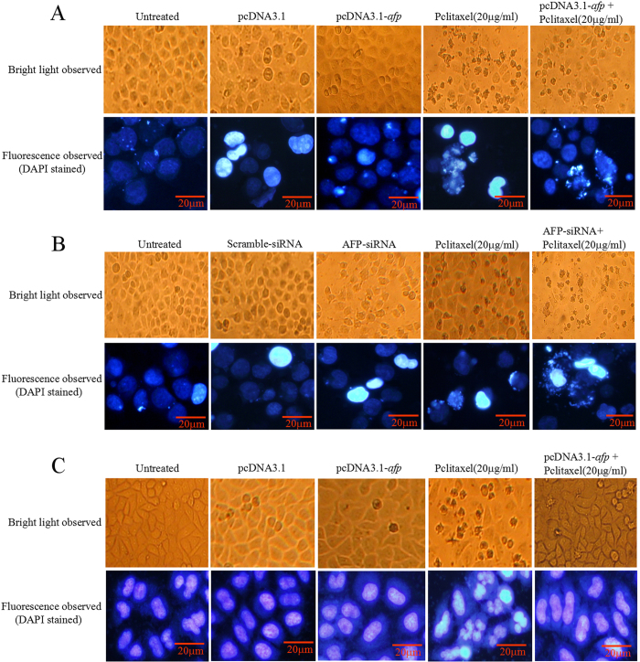
Figure 3. Effects of AFP on paclitaxel inducing apoptosomes in human hepatoma cells and L-02 cells in vitro.
(A) HLE cells were treated with paclitaxel at a concentration of 20 μg/ml and transfected with pcDNA3.1-afp vectors for 24 hrs followed by treatment with paclitaxel (20 μg/ml) for 24 hrs. The morphological changes in HLE cells were observed by microscopy. The cytoblasts of HLE cells were stained with DAPI and observed by fluorescent microscopy. B, Bel 7402 cells were treated with paclitaxel at a concentration of 20 μg/ml and transfected with AFP-siRNA vector for 24 hrs followed by treatment with paclitaxel (20 μg/ml) for 24 hrs. The morphological changes in Bel 7402 cells were observed by microscopy. The cytoblasts of Bel 7402 cells were stained with DAPI and observed by fluorescent microscopy. C, L-02 cells were treated with paclitaxel at a concentration of 20 μg/ml and transfected with pcDNA3.1-afp vectors for 24 hrs followed by treatment with paclitaxel (20 μg/ml) for 24 hrs. The morphological changes in L-02 cells were observed by microscopy. The cytoblasts of L-02 cells were stained with DAPI and observed by fluorescent microscopy. The images were representative of at least three independent experiments.

