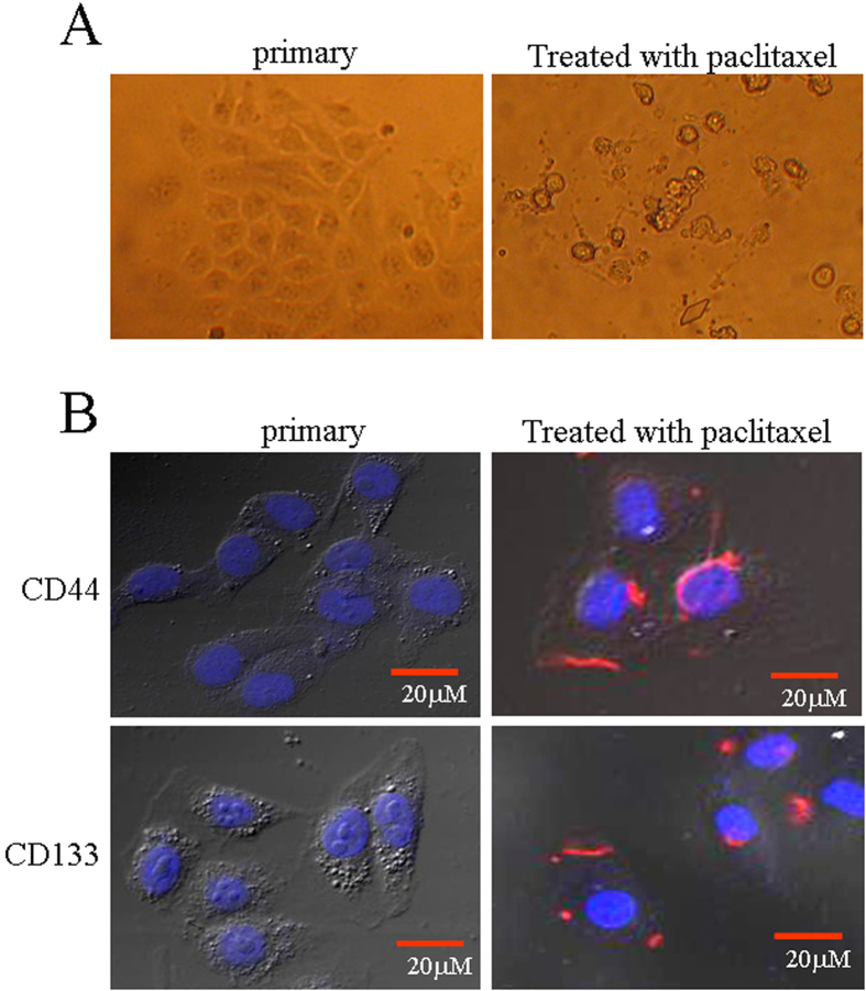
Figure 5. Expression of stemness markers in paclitaxel-resistant Bel 7402 cells.
(A) Bel 7402 cells were treated with paclitaxel (20 μg/ml) for 48 hrs, and the morphologic changes in the cells were observed by bright-light microscopy. (B) Bel 7402 cells were consecutively treated with paclitaxel (20 μg/ml) for 7 d, and the cells were washed with PBS three times. The expression of the stemness markers CD44 and CD133 in the cells was observed by laser confocal microscopy; the red staining shows the location of the markers. The images are representative of at least three independent experiments.

