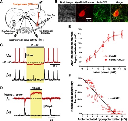
Figure 6.
Optogenetic inhibition of rhythmically active Arch-expressing pre-BötC inspiratory glutamatergic neurons in vitro. A, Overview of experimental in vitro neonatal mouse rhythmic slice preparation with unilateral pre-BötC laser illumination (2-15 mW). NA, Nucleus ambiguus; V4, fourth ventricle. B, Two-photon single-optical plane images of pre-BötC glutamatergic neuron targeted for whole-cell recording, showing a Dodt structural image, VgluT2-Cre-mediated tdTomato labeling, and Arch-GFP expression (coexpression confirmed in merged image). C, Current-clamp recording from pre-BötC inspiratory neuron in B illustrates inspiratory bursting synchronized with integrated inspiratory hypoglossal activity (∫XII). The membrane potential was hyperpolarized by ∼10 mV at 10 mW laser power. This unilateral illumination was sufficient to reduce the frequency and amplitude of ∫XII. D, Under voltage-clamp, the same neuron as in C exhibited rhythmic inward synaptic currents synchronized with ∫XII, whereas laser illumination (10 mW) induced outward currents of ∼200 pA. E, Summary data showing Arch-mediated hyperpolarization in VgluT2-expressing pre-BötC inspiratory neurons (n = 22) was laser power dependent. After blocking fast glutamatergic synaptic transmission with CNQX, laser-induced hyperpolarization of VgluT2-positive pre-BötC inspiratory neurons (n = 6) was also laser power dependent, which was not significantly different from the cases with the synaptically coupled network (unpaired t test, p = 0.938). F, Linear correlation between laser-induced hyperpolarization of VgluT2-positive pre-BötC inspiratory neurons (n = 22) and XII inspiratory frequency, showing the monotonic reduction of frequency as a function of membrane hyperpolarization. Data are represented as the mean ± SD.

