
Figure 3. Multi-scale visualization of prostate surface images from the macro-scale to the micro-scale.
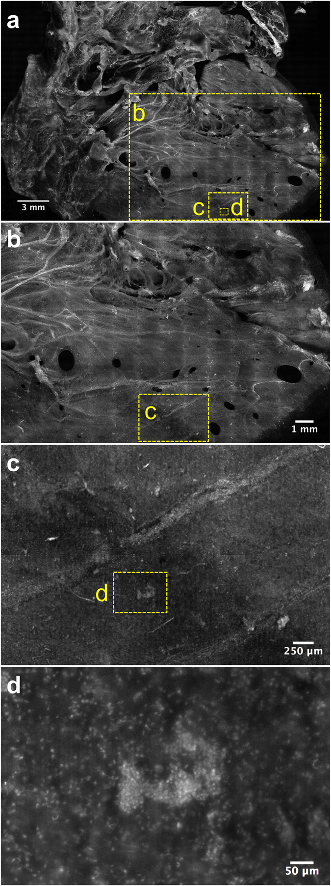
(a) VR-SIM image of the 6.5 cm2 right lateral surface of Case 6, (b) Zoom of the large dashed yellow box area indicated in (a). Features such as the neurovascular bundles (bright spindly features) and smooth prostatic pseudocapsule/fascia are apparent. (c) Zoom of the dashed yellow box in (b). The image is marked by a single neurovascular bundle extending across the field of view. A bright feature (d) is enclosed by a dashed yellow box, corresponding to a single prostate gland. Individual rounded nuclei are clearly resolved.