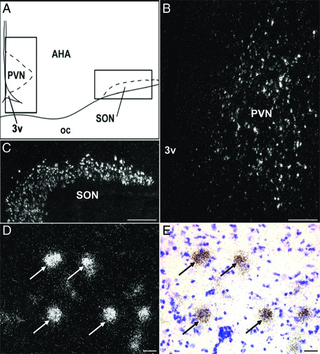
Figure 2.
KOR mRNA in PVN and SON. Camera lucida drawing (A) displaying areas (solid box) depicted in dark-field photomicrographs (B and C) of a representative hypothalamic section hybridized with antisense probe to ovine KOR mRNA. Cells expressing KOR were observed in the PVN (B) and SON (C). High-power dark- (D) and bright-field (E) photomicrograph of PVN cells expressing KOR (arrows). Scale bar (B and C) 500 μm; (D and E) 100 μm. oc, optic chiasm; 3v, third ventricle.

