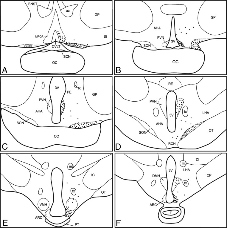
Figure 4.
Camera lucida drawings illustrating representative distribution of KOR-ir cells in the POA (A), SON (B–D), PVN (B–D), AHA; anterior hypothalamic area (B–D), retrochiasmatic area (RCH; D), VMH (E) and DMH (F) nuclei of the hypothalamus; and ARC (E and F). Each black dot represents approximately 10 KOR-ir cells. The distribution of KOR-ir was identical on both sides of the POA and hypothalamus and is shown unilaterally to allow visualization of labels. ac, anterior commissure; BNST, bed nucleus of the stria terminalis; CP, cerebral peduncle; fx, fornix; GP, globus pallidus; ir, infundibular recess; mt, mammillary tract; OC, optic chiasm; OT, optic tract; OVLT, organum vasculosum of lamina terminalis; PE, periventricular nucleus; PT, pars tuberalis of the adenohypophysis; RE, reticular nucleus; SCN, suprachiasmatic nucleus; SI, substantia innominata; ZI, zona incerta; 3v, third ventricle.

