Fig. 1.
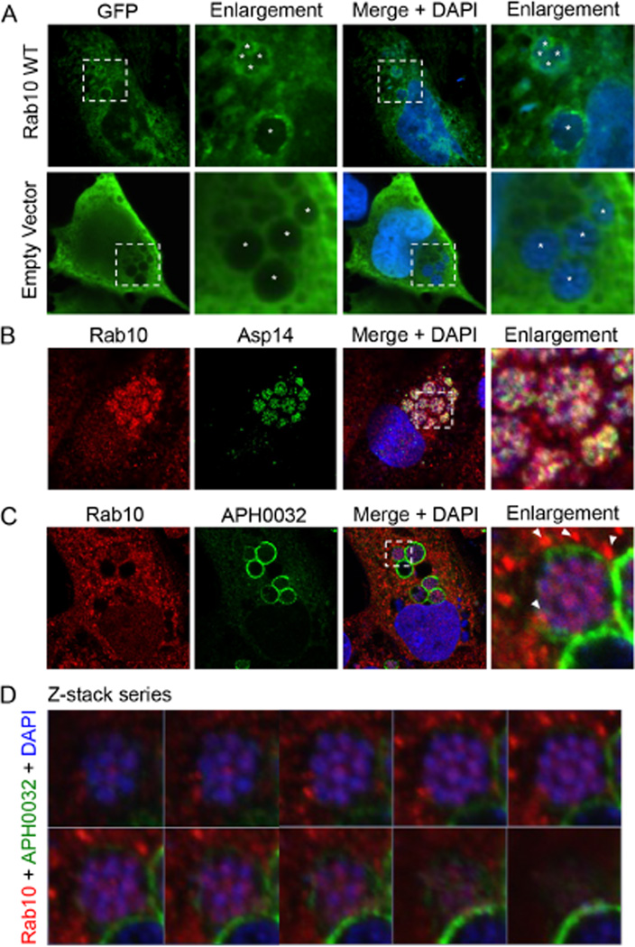
Endogenous and ectopically expressed Rab10 localize to the AVM and with intravacuolar A. phagocytophilum organisms.
A. GFP-Rab10 localizes to the AVM and with intravacuolar bacteria. RF/6A cells expressing GFP-Rab10 were infected with A. phagocytophilum and visualized by LSCM. Asterisks denote bacterial vacuoles.
B. Endogenous Rab10 localizes with A. phagocytophilum surface protein Asp14. A. phagocytophilum-infected RF/6A cells that had been screened with antibodies against Rab10 and Asp14 were examined by confocal microscopy.
C, D. Z-stack series and 3D rendering show endogenous Rab10 associated with A. phagocytophilum organisms within the ApV. A. phagocytophilum-infected RF/6A cells were screened with antibodies against Rab10 and AVM protein APH0032 and visualized by confocal microscopy. (C) Representative infected host cell that was used for Z-stack analysis. Note the localization of punctate Rab10 labelling at the AVM (arrow heads) and within the ApV associated with A. phagocytophilum organisms. (D) Z-stack series showing individual, successive focal planes. (A–C) The regions in the merge + DAPI panels that are demarcated by hatched-line boxes indicate the regions that are magnified in the enlargement panels. The enlargement panel in C was analysed by Z-stack in (D) (A–D) Host cell nuclei and bacterial DNA were stained with DAPI (blue). Results shown are representative of three experiments with similar results.

