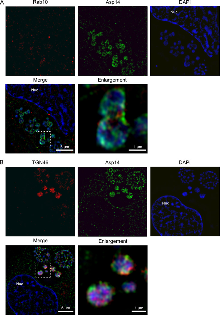
Fig. 4.
Structured illumination microscopic analyses confirm that Rab10-positive and TGN46-positive vesicles are present in the ApV proximally associated with intravacuolar A. phagocytophilum organisms.
A, B. A. phagocytophilum-infected RF/6A cells that had been screened with antibodies against (A) Rab10 and Asp14 or (B) TGN46 and Asp14 were examined by SIM. The regions in the merge + DAPI panels that are demarcated by hatched-line boxes indicate the regions that are magnified in the enlargement panels. Host cell nuclei and bacterial DNA were stained with DAPI (blue). Results shown are representative of three experiments with similar results. Nuc, host cell nucleus.

