Fig. 5.
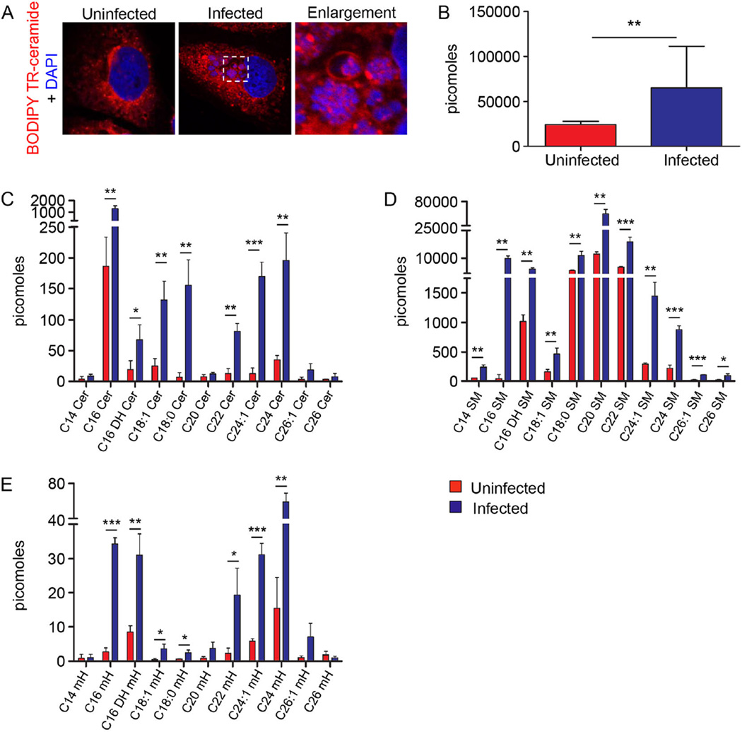
Sphingolipids are delivered into the ApV and incorporated by A. phagocytophilum.
A. BODIPY TR-labelled sphingolipids localize with intravacuolar A. phagocytophilum organisms. BODIPY TR ceramide was added to uninfected and A. phagocytophilum-infected cells and examined by LSCM. Host cell nuclei and bacterial DNA were stained with DAPI (blue). Results are representative of three experiments.
B–E. A. phagocytophilum incorporates host cell sphingolipids. Host cell-free A. phagocytophilum DC organisms recovered from the media of infected HL-60 cells were subjected to UPLC-ESI-MS/MS to detect total sphingolipids (B), ceramides (Cer) (C), sphingomyelins (SM) (D) and monohexosyl sphingolipids (mH) (E). Media from uninfected HL-60 cells was processed and examined in parallel. Results are the mean ± standard deviation of triplicate samples and are representative of three biological replicates with similar results. Statistically significant (*P ≤ 0.05; **P ≤ 0.01; ***P ≤ 0.001) values are indicated.

