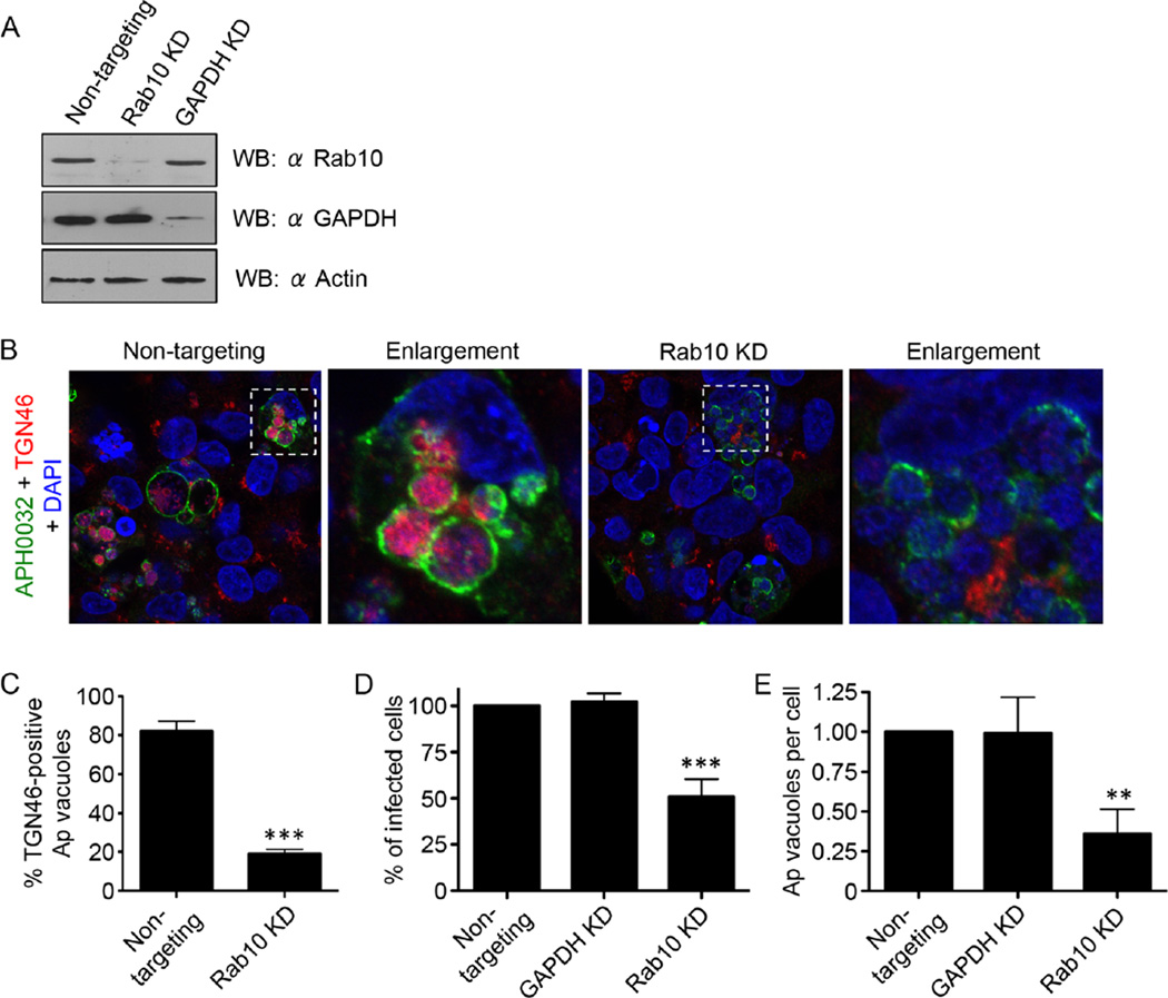
Fig. 7.
Knockdown of Rab10 markedly reduces both TGN vesicle delivery into the ApV and A. phagocytophilum infection. HEK-293T cells were treated with Rab10-targeting, non-targeting or GAPDH-targeting siRNA for 72 h.
A. Western blot showing knockdown of Rab10 or GAPDH after appropriate siRNA treatment.
B–D. After siRNA treatment, the cells were infected with A. phagocytophilum (Ap) for 48 h and processed for confocal microscopy. (B and C) Rab10 knockdown markedly inhibits translocation of TGN vesicles into the ApV. Rab10-targeting and non-targeting siRNA-treated infected cells were screened with antibodies against APH0032 and TGN46 and stained with DAPI (blue). (B) Representative confocal micrographs. Regions that are demarcated by hatched-line boxes are magnified in the corresponding enlargement panels. (C) Percentage ± standard deviation (SD) of TGN46-positive ApVs in non-targeting siRNA-treated or Rab10-targeting siRNA-treated infected cells. (D and E) Rab10 knockdown (KD) significantly reduces A. phagocytophilum infection. siRNA-treated infected cells were screened with an antibody against A. phagocytophilum P44 to denote bacteria and examined by immunofluorescence microscopy to determine the percentage ± SD of infected cells (D) and mean number ± SD of ApVs per cell (E). Statistically significant (**P ≤ 0.01; ***P ≤ 0.001) values are indicated. Data presented in each panel are representative of three experiments with similar results.

