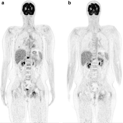
Fig. 2.

Coronal 18F-FDG PET-CT scans done prior to (a) and after (b) 3 cycles of pembrolizumab. The markedly increased FDG uptake in the right side and adjacent soft tissues of T12, in the left ischium and in one of the right middle lobe pulmonary metastases are shown. Post-treatment the FDG avidity in the bony lesions is much reduced and the right middle lobe lesion had completely resolved
