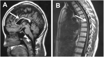
Fig. 3.

Sagittal brain and thoracic spinal MRI image in index patient (II:3). a: Brain MRI shows thinning of corpus callosum, emphasized in the anterior part with a “beaked” shape (arrow). Ther is no obvious periventricular or deep cerebellar white matter lesions and no obvious cerebral or cerebellar atrophy. b: Throracic spinal MRI shows thinning of the thoracic spinal cord with volume loss but no obvious signal abnormalities in the cord
