Fig. 1.

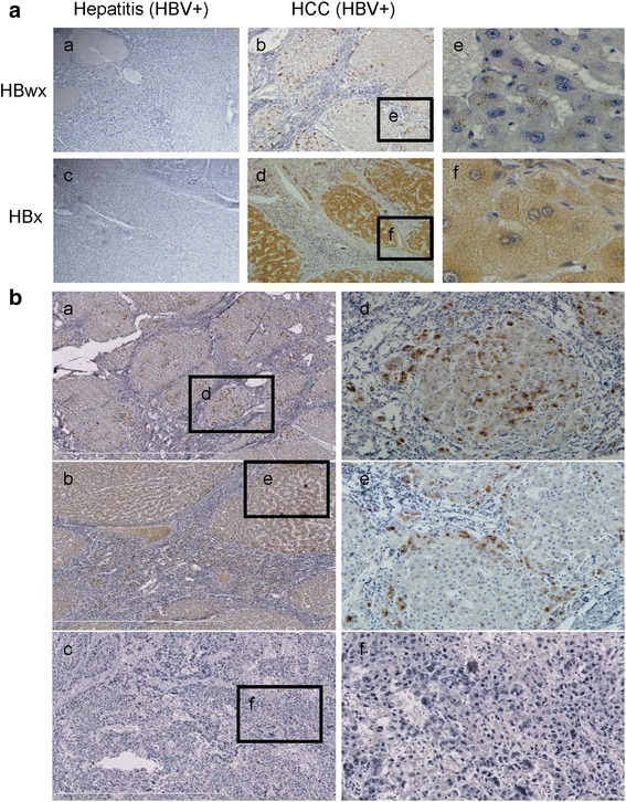
Subcellular distribution of HBwx and HBx and characteristics of HBwx distribution in HBV-associated HCC specimens detected by immunohistochemistry. a HBwx immunoreactivity was mainly observed in the nucleus and in the cytoplasm surrounding the nuclear membrane (e). Immunohistochemical staining of HBx revealed diffuse positivity in the hepatocytes, especially in cytoplasm (f) (a-d, original magnification, ×100) (e-f, original magnification, ×400). b More intense and widespread HBwx immunoreactivity was observed in the small tumor masses (a, d) or the peripheral parts of the large tumor masses near the portal area (b, e); No HBwx immunoreactivity was observed in tumor cells of poorly differentiated hepatocellular carcinoma (c, f) (a-c, original magnification, ×100) (d-f, original magnification, ×200)
