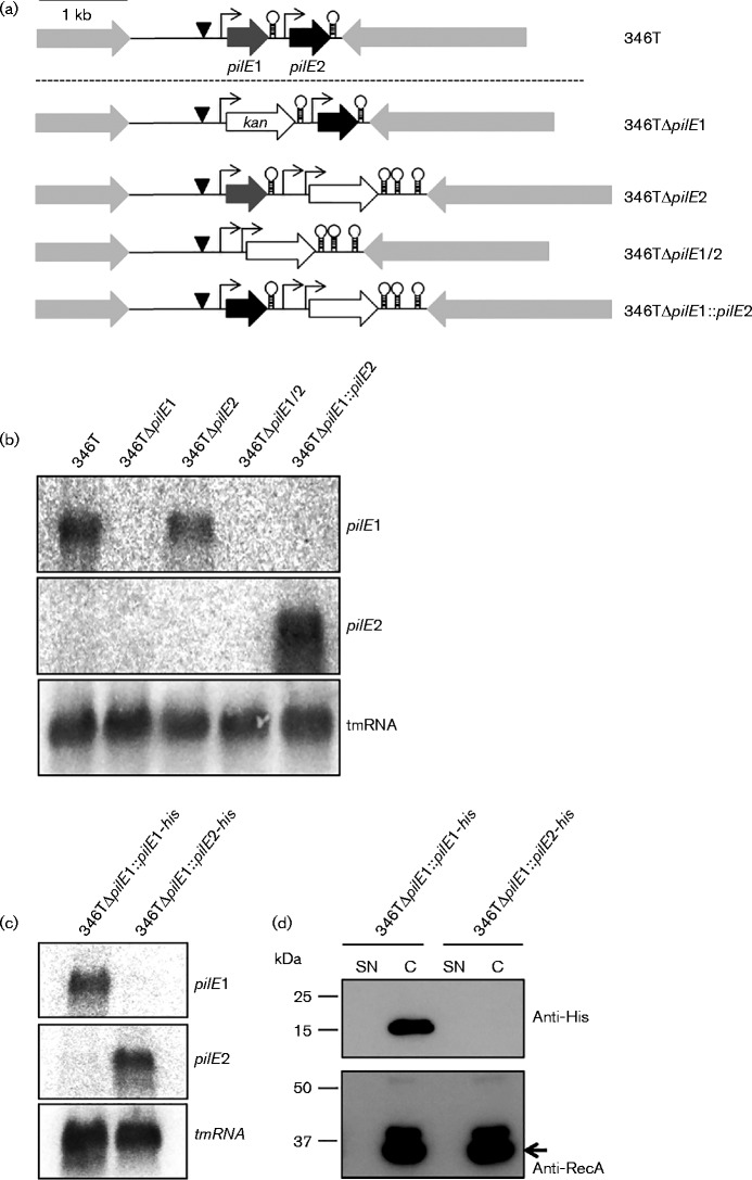
Fig. 2.
Analysis of pilE1 and pilE2 transcript and protein expression in N. cinerea CCUG 346T. (a) Schematic representation of the WT pilE locus in N. cinerea 346T and isogenic mutants used to analyse transcript expression. Putative promoters are indicated by arrows (σ70) and triangles (σ54), and Rho-independent terminators by stem–loops. A kanamycin resistance cassette with promoter and bidirectional rrnB terminators was used for the construction of mutants, except for 346TΔpilE1, in which only the ORF was used. Bar, 1 kb. (b) Northern blotting of total RNA from N. cinerea 346T WT and mutants. pilE1 transcript was detected in both the WT and 346TΔpilE2. pilE2 transcript was undetectable in the WT under the conditions tested but was detected when expressed from the pilE1 promoter, in 346TΔpilE1 : : pilE2. tmRNA was used as loading control. (c) Northern blot analysis of total RNA from 346TΔpilE1 : : pilE1-his and 346ΔpilE1 : : pilE2-his. pilE1 and pilE2 transcripts were detected, respectively. tmRNA was used as loading control. (d) Detection of His-tagged PilE1 and PilE2 in supernatant (SN) and cell extracts (C) from strains 346TΔpilE1 : : pilE1-his and 346TΔpilE1 : : pilE2-his. RecA detection was used as loading control. No signal was obtained for PilE2.

