Fig. 5.
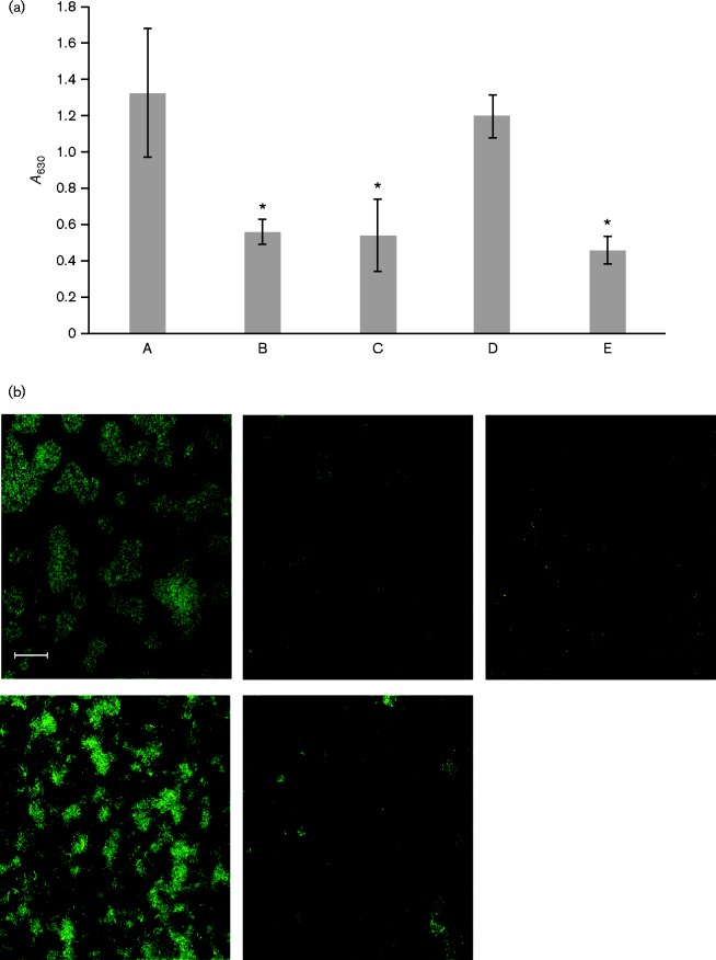
Quantification and confocal microscopy of biofilm formation of strains of A. actinomycetemcomitans. (a) Quantification of biofilm mass using a crystal violet assay. A. actinomycetemcomitans strains were grown as biofilms in glass-bottomed dishes and stained with SYTO 9. A series of Z-stack images was generated with a Zeiss LSM 510 META confocal microscope, and biofilm area was quantified using ImageJ. Asterisks indicate significant difference from the WT fimbriated strain (ANOVA with Dunnett's post-test, P < 0.05). A, Fimbriated clinical isolate (VT1257); B, WT (VT1169); C, morC mutant (VT1650); D, WT/pKM2/flp1-tadV (KM611); E, morC mutant/pKM2/flp1-tadV (KM612). Results are shown as means ± sd. (b) Representative fields of confocal microscopy of biofilms formed by the strains in (a). Upper left: Fimbriated clinical isolate (VT1257); middle: WT (VT1169; upper right: morC mutant (VT1650); lower left: WT/pKM2/flp1-tadV (KM611); lower right: morC mutant/pKM2/flp1-tadV (KM612). Bar, 20 μm.

