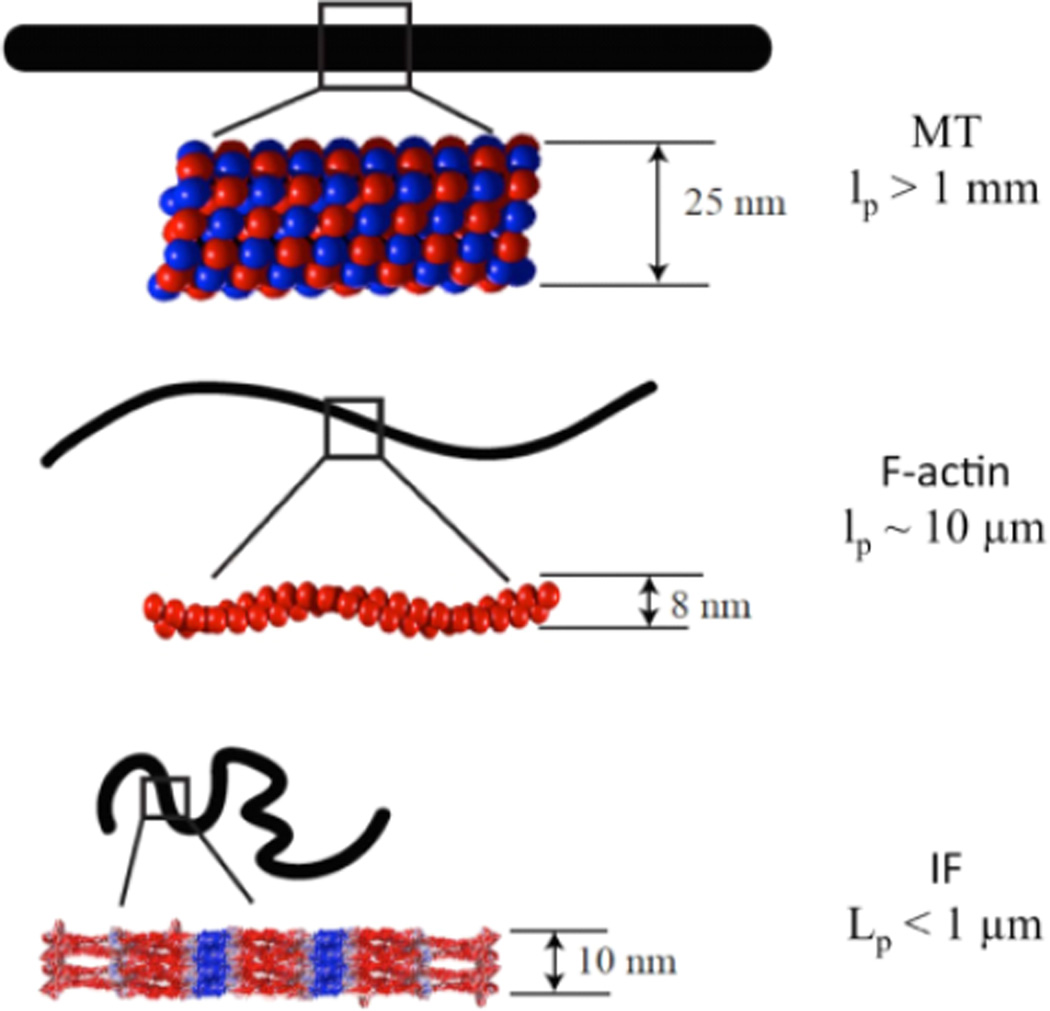
Figure 1.
Schematic diagram of approximate diameter, subunit packing and filament configuration of each of the three cytoskeletal polymer types: microtubules (MT), F-actin, and intermediate filaments (IF). The black filament outline represents the configuration of each filament in solution at 37°C due to the thermal fluctuations acting on 10 micron long filaments with the persistence lengths lp listed on the right.

