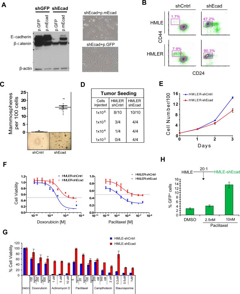
Figure 1. Mesenchymally transdifferentiated breast epithelial cells have increased numbers of CSCs and are drug resistant.
(a) Western blotting for E-cadherin, β-catenin and β-actin in HMLER cells expressing either GFP (shGFP) or the human ECAD gene (shEcad). Stable introduction of a murine ECAD gene (p.mEcad) but not GFP (p.GFP) results in re-expression of E-cadherin protein and reversal of EMT-associated morphology. (b) FACS with CD24 and CD44 markers; Percentage of the CD44+/CD24-subpopulation is indicated. (c) Mammosphere formation assays and (d) tumor-seeding with HMLERshCntrl and HMLERshEcad breast cancer cells. (e) Proliferation curves of HMLER-shCntrl and HMLER-shEcad cells grown in culture. Viable cells were counted by Trypan Blue dye-exclusion. (f) Dose-response curves of HMLERshEcad and HMLERshCntrl breast cancer cells treated with doxorubicin or paclitaxel. (g) Viability of immortalized, non-tumorigenic breast epithelial cells (HMLE shCntrl) and cells induced through EMT (HMLEshEcad) treated with various chemotherapy compounds (h) Proportion by FACS of GFP-labeled HMLEshEcad cells following paclitaxel treatment when mixed with control cells (HMLE) cells.

