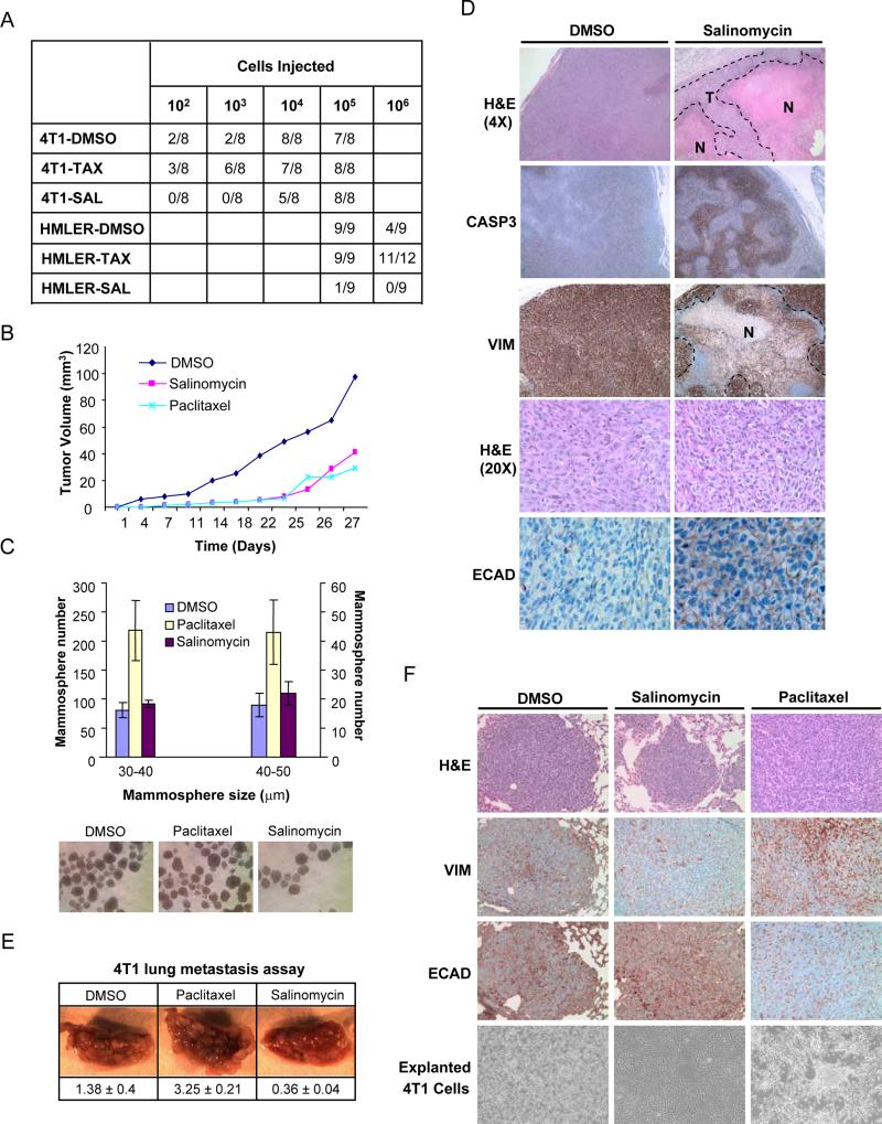
Figure 5. Effects of salinomycin and paclitaxel treatment on tumor seeding, growth and metastasis in vivo.
(a) Tumor-seeding ability of HMLER and 4T1 breast cancer cells treated with salinomycin, paclitaxel or DMSO. (b) SUM159 tumor-growth curves of compound-treated mice. (c) Quantification of tumorsphere-forming potential (diameter between 20-50μm was evaluated) of cancer cells isolated from dissociated SUM159 tumors from compound-treated mice. Images of tumorsphere cultures are shown. (d) Histological analysis of tumors from salinomycin- or vehicle-treated mice. Shown are H&E, caspase-3, human-specific vimentin and E-cadherin staining. (e) Tail-vein injection of 4T1 cancer cells, pre-treated with paclitaxel, salinomycin, or DMSO. Lung images shown were captured at 1.5X magnification. Values are shown below the images as the mean and standard error for lung burden in each treatment group. (f) H&E, vimentin and E-cadherin staining of lung nodules from compound-treated 4T1 breast cancer cells. Also shown are images of cultured 4T1 cells explanted from lung nodules.

