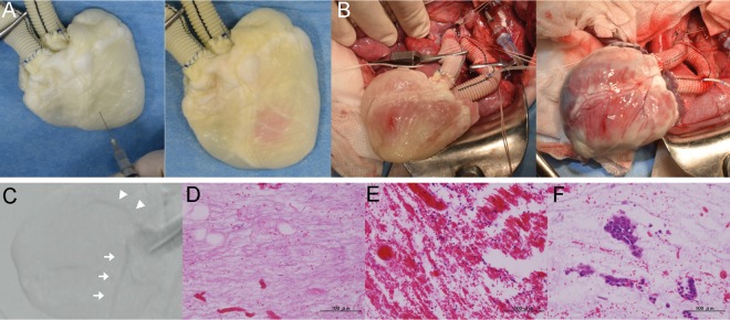
Figure 6:
Heterotopic transplantation of the recellularized heart with the muscular injection method. (A) Representative photography of the muscular injection of pig mesenchymal stem cells in the decellularized heart. (B) Operative findings of the scaffold, before (left) and after (right) de-clamping. (C) Digital subtraction angiography of the coronary artery. White arrow: left coronary artery. Arrowhead: right coronary artery. (D–F) Haematoxylin and eosin staining of the recellularized transplanted heart. Nearly intact ventricular muscle at the apex (D). Thrombosis in the parenchymal space (E). Injected porcine mesenchymal stem cells (F). Scale bar: 100 μm.

