Figure 5.
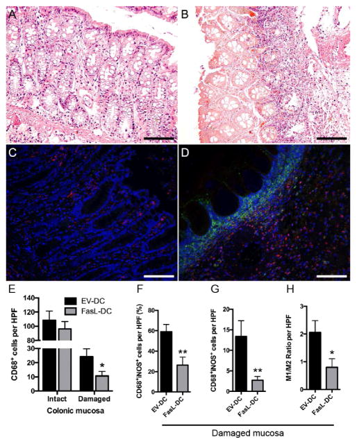
Adoptive transfer of dendritic cells expressing FasL (FasL-DCs) decreases the proportion of colonic proinflammatory macrophages. Representative micrographs of hematoxylin and eosin staining depicting the histology of intact (A) and damaged (B) colonic mucosa from colitic rats (400×). Representative micrographs of double immunofluorescent staining for CD68 (red) and inducible nitric oxide synthase (iNOS, green) of intact (C) and damaged (D) colonic mucosa from colitic rats (400×). (E) Number of CD68+ cells per high-power field (HPF, 400×) of colonic tissue from rats treated with FasL-DCs or dendritic cells transfected with an empty vector (EV-DCs). (F) quantification of CD68+iNOS+ cells per HPF of colonic tissue from rats treated with FasL-DCs or EV-DCs, expressed as a percentage of total CD68+ cells. (G) Number of CD68+iNOS+ cells per HPF of colonic tissue from rats treated with FasL-DCs or EV-DCs. (H) M1/M2 ratio (calculated CD68+iNOS+/CD68+iNOS−) per HPF of colonic tissue from rats treated with FasL-DCs or EV-DCs. n=14–15 HPFs for intact mucosa, 15–17 HPFs for damaged mucosa. * p<0.05, ** p<0.01 vs EV-DC group. Scale bars = 100 μm.

