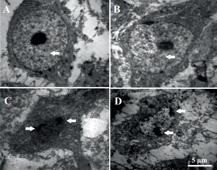
Figure 5.
Injection of Aβ into the frontal cortices induced ultra-structural changes in CA1 pyramidal neurons. Electron microscopic photomicrographs from CA1 pyramidal neurons of control (A) and sham rats (B), in which arrows show oval nuclei with evenly dispersed chromatin and clear nucleoli in the center of nucleus. Electron microscopic photomicrographs from CA1 pyramidal neurons of Aβ (C) and Aβ+melatonin (D) treated rats, respectively, in which arrows indicate nucleolemma invaginations and chromatin aggregation.

