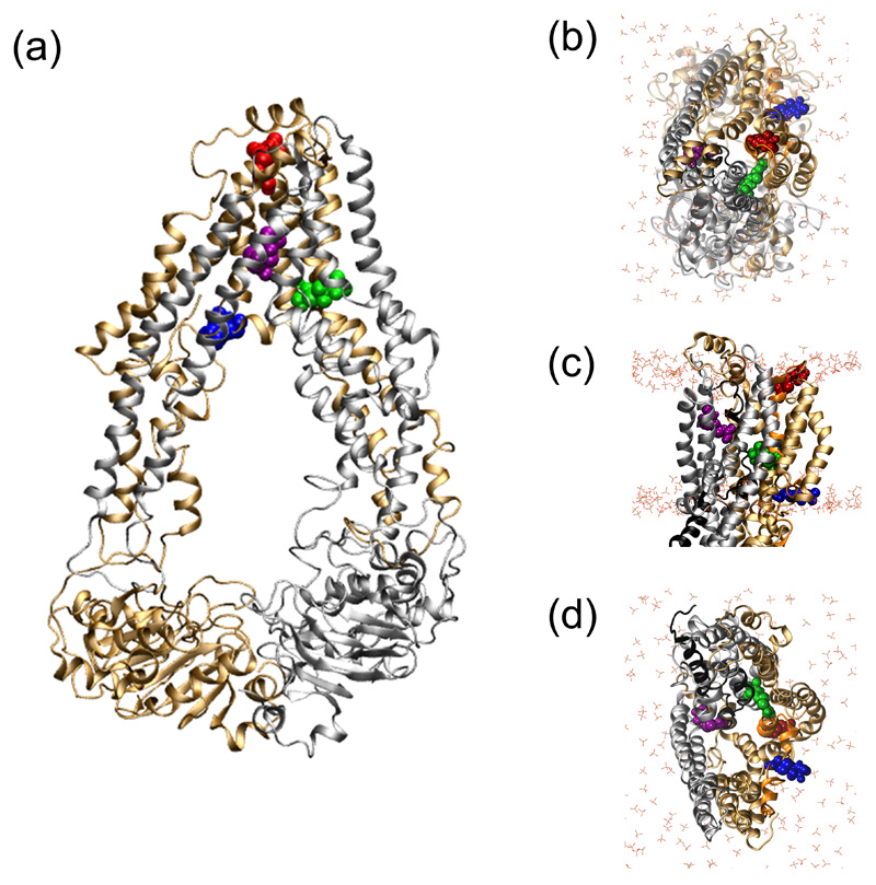
Figure 4. Location of the four cysteine-linked MTSL probes.
The location and orientation of the cys-MTSL moiety incorporated into specific positions in the MD equilibrated membrane-embedded ABCB1 (starting conformation: adapted from PDB 3G5U-a) are shown in panels (a-d). The N- and C-terminal domains of ABCB1 are in bronze and grey ribbons, respectively. MTSL labelled 331C (red), 343C (green), 354C (blue) and 980C (purple) are shown in spacefill. The POPC headgroups are in liquorice with CPK colouring. The NBDs have been removed from the structures in panels (b-d) for clarity.
a) Structure of the MD equilibrated ABCB1 (PDBid: 3GU-a) viewed along the plane of the membrane.
b) View of MD equilibrated ABCB1 from the extracellular side of the membrane.
c) Side profile of MD equilibrated ABCB1.
d) The MD equilibrated ABCB1 TMD cavity, viewed from the intracellular side of the membrane.

