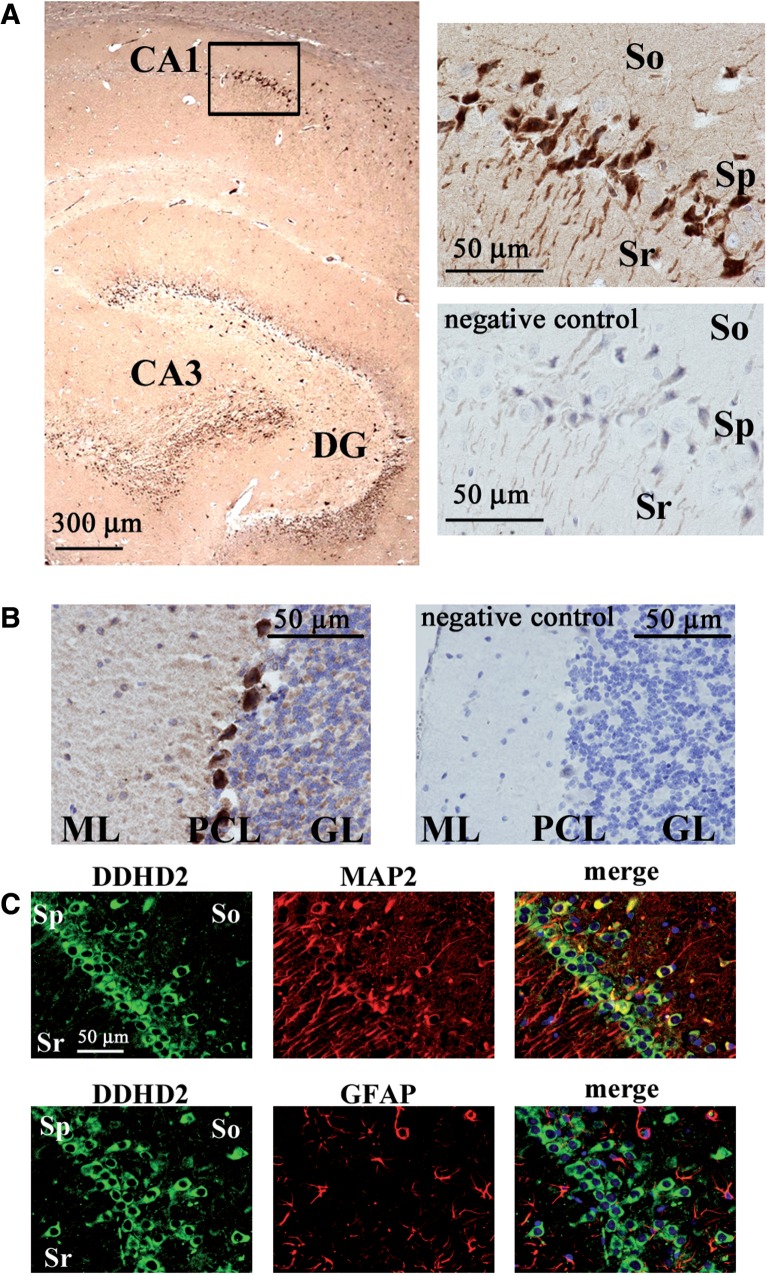
Fig. 10.
Immunostaining of DDHD2 in rat brain. (A) The distribution of DDHD2 is shown using a sagittal section of the hippocampus in a rat brain. DDHD2 was stained by the anti-DDHD2 antibody with DAB (brown). Nuclei were stained by with hematoxylin (blue). The rectangular area is magnified in the upper right panel. The lower right panel is from a negative control experiment using the excessive peptides used for the immunization (16 µg/ml) to absorb primary antibody. (B) The distribution of DDHD2 is shown using a sagittal section of the cerebellum in a rat brain. The right panel is from a negative control experiment. ML, molecular layer; PCL, Purkinje cell layer; GL, granule cell layer. (C) The distribution of DDHD2 is shown using a coronal section of a rat brain. Double staining of DDHD2 and MAP2 (upper panels) or DDHD2 and GFAP (lower panels) at the CA1 region in the hippocampus shows that DDHD2 is localized in the cell body of hippocampal neurons. Sr, stratum radiatum; So, stratum oriens; Sp, stratum pyramidale. Data are representative of three independent experiments.

