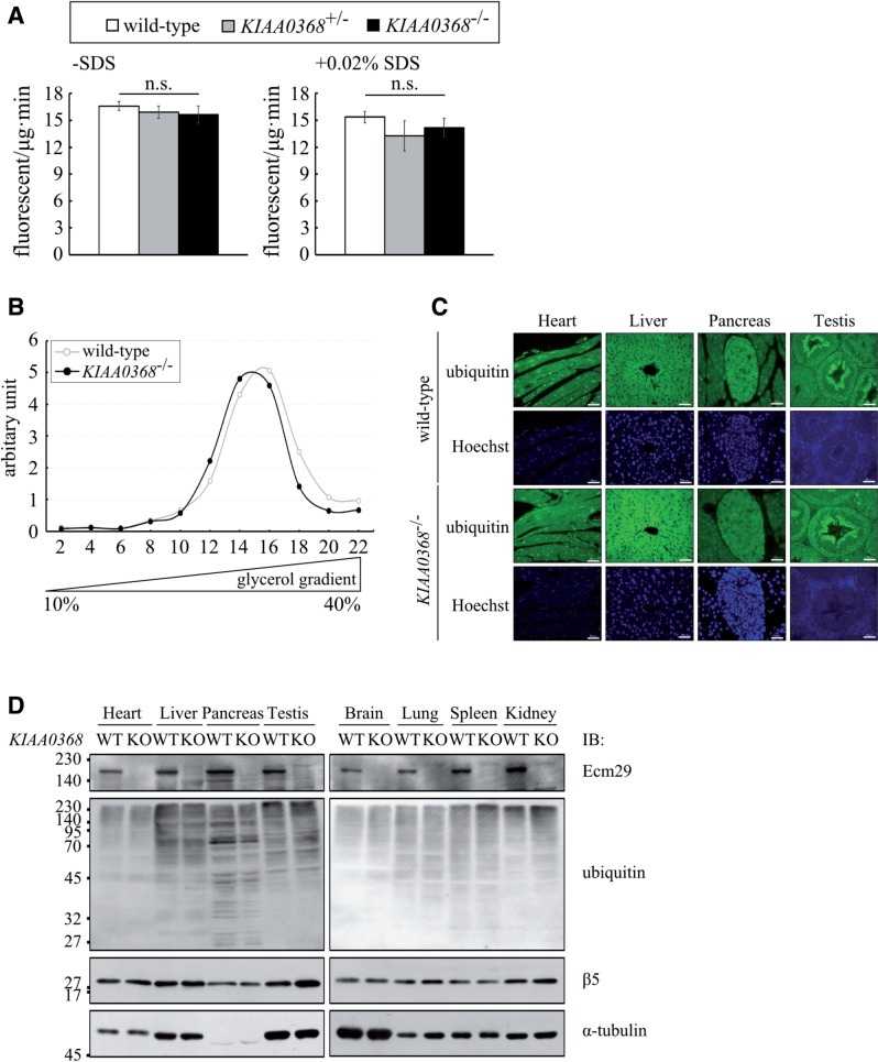
Fig. 2.
Proteasomal peptidase activities in KIAA0368-deficient mice. (A) Peptidase activities of testes lysates. Hundred micrograms of testes lysates from wild-type, heterozygous or homozygous mice for KIAA0368, were incubated with fluorescent substrates in the presence or absence of 0.02% SDS. The bar charts represent the mean ± SD of fluorescence/μg/min. LLVY; chymotrypsin-like activity. White bars; wild-type, grey bars; KIAA0368+/-, black bars; KIAA0368−/−. n.s.; not significant by student’s t-test. (B) Peptide hydrolysis activities of sedimentation velocity fractions from wild-type or KIAA0368-deficient testes lysates. 1 mg of testis lysates were fractionated by glycerol density gradient centrifugation (10–40% glycerol from 1 to 22 fractions). Suc-LLVY-AMC was used to measure the peptidase activities. The relative peptidase activities (fluorescence/min) normalized to the first fraction are shown. (C) Tissue sections from wild-type or KIAA0368-deficient mice were immunostained with ubiquitin antibody and counterstained with Hoechst 33342. Scale bars indicate 50 µm. (D) Tissue blots of wild-type (WT) and KIAA0368-deficient mice (KO). 50 μg of heart, liver, pancreas, testis, brain, lung, spleen and kidney lysates were subjected to immunoblot analyses using antibodies against Ecm29, Multi-ubiquitin, β5 and α-tubulin.

